Click Here for More Images from iStock- 15% off with coupon 15FREEIMAGES 
Ultrasound upper abdomen showing flow in common bile duct  after use color Doppler.
Latina doctor checking a patient's arm in the clinic's office.
Hospital monitor displaying vital signs: heart rate, blood pressure, oxygenation, temperature, and end-tidal CO2, crucial for patient care and health assessment
Computational tomography image of the abdominal and pelvic regions.
full abdomen ultrasound image aka diagnostic sonogram
abstract composition on a theme of Science, Technology and research
1970s Slide Rule
Matured Asian medical doctor examining lungs X-ray negative film on white background
Stock market chart. Treading board. Profit and money. Financial and business graph. Buy and sell concept. Stock market volatility 3d illustration.
CT SCAN of Chest and Abdomen Coronal and sagittal view with injection contrast media for diagnosis chest and abdomen disease.
Monitoring the patient's wellbeing
ct scan step set of neck coronal view
Computer control equipment manufacturing parts
Heart ultrasound image on a computer screen.
Hospital
Doctors observing the progress of a cardiopulmonary stress test taken by the male athlete riding a bicycle ergometer. Young adult man having a VO2 test with a VO2 mask on his face, electrocardiogram pads attached, computer recording, indoor bicycle. Healthcare and Medicine concept.
Skan of western blotting film with an image of four membranes subjected to antibodies recognizing different protein species visible as black stops or blots
Iron ruler on a white background. Stainless steel ruler. Stationery and work tools made of metal steel. Selective focus, isolated on white background
Full size digital screening mammogram of a new breast tumor in the retroareolar region of the right breast causing nipple retraction. An ultrasound guided biopsy was performed diagnosing invasive ductal carcinoma. This is the most common type of breast cancer, accounting for nearly 80% of cases. Breast cancer treatment has come a long way in the last few decades, seeing more funding than any other human disease. The first step to a cure is catching it early with screening mammograms such as this case so that the tumor can be removed before it spreads (metastasizes) out of the breast.
normal abdomen CT scan
Mammogram
Mediastinal view of a hamartoma tumor on the left lung. Medical themes
Computational tomography image of the abdominal and pelvic regions.
Rear view of male Japanese doctor examining patient x-rays in illuminated light box at Tokyo hospital.
Metal Ruler isolated on white background.
MRI of the prostate gland, revealing an enlarged size, aids in diagnosing tumors, guiding treatment decisions, and monitoring prostate health.
Cardiogram Chart (ECG). Close-up. Hospital.
Ophthalmic test - OCT optical coherence tomography measurement. SLO Scan view of the macula in retina with vessels
Ophthalmologist modern clinic
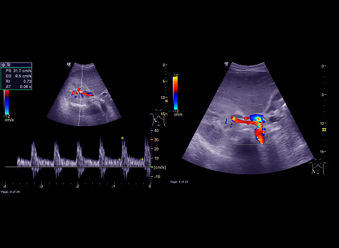
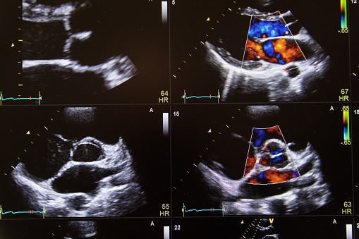
Free Images: "bestof:PLAX Mmode.jpg Echocardiogram in the parasternal long-axis view showing a measurement of the heart's left ventricle Uploadet by Kjetil Lenes who made the"
PLAX Mmode.jpg
Ventricular Septal Defect.jpg
Echokardiogram von Atriumseptumdefekt (Ostium secundum).jpg
Echocardiogram 4chambers.jpg
Stent tekst.jpg
PDA Coil.png
Coarctation and PDA.png
Ventricular septal defect (schematic drawing).png
Aortic divisions.png
Aortic arch with vessels.png
Schematic drawing of various types of atrial septal defect.png
Foramen ovale.png
Flow in pulmonic valve.jpg
Schematic mollusc.jpg
Terms of Use   Search of the Day