Click Here for More Images from iStock- 15% off with coupon 15FREEIMAGES 
DNA close-up, DNA Molecule Model, DNA Strands, DNA animation
Phospholipid bilayer membrane structures that make up living cells and their contents. This is an image of membrane proteins in the membrane.\nThis image, which looks like a plastic model, evokes biology and biochemistry. It can be used as a cover for magazines, textbooks and presentation materials.\nThis image was not created by AI.
orange: RNA primer; red: RNA template; light blue: proofreading riboexonuclease; mid blue: helicase; dark blue: RNA directed RNA polymerase; violet: non structural proteins (NSPs).
Endosomes - 3d rendered image of Endosomes are membrane-bound organelles. They are parts of endocytic membrane transport pathway.  Endomembrane system in cells. Bilayer Lipid Membrane. Abstract medical illustration. Microscopic view. Microbiologic research concept.
3D cartoon and Gaussian surface models, chain id color scheme, PDB 3omv, white background
A ball and stick + ribbons model of beta-secretase - a protein that is important in forming healthy peripheral nerve cells, but is also involved in the onset of Alzheimer's disease.
Proteasomes are molecular machines for breaking down proteins called proteolysis, only target proteins that have been marked for destruction called ubiquitin to the target protein, 3d rendering
Covid-19 Coronavirus Variants Omicron Delta Gamma Beta Alpha Pandemic Virus Disease Mutation
DNA mutation, a mutation is a change in a DNA sequence, mutations can result from DNA copying mistakes made during cell division.
Yellow Chana Dal or Split Gram Lentils or Pulses as ingredients toward Indian and South Asian Cuisine
The LDL-receptor (red) is a membrane protein found in almost every human cell. It binds to the apoprotein B100 from LDL-particles and mediates their internalization (endocytosis) in the cell. This occurs in almost all cells but mainly in the liver where it removes about 70 % of LDL from the circulation.
Cells in the body examined by endoscope
3D cartoon and Gaussian surface models, PDB 2j0D, white background.
Endosomes - 3d rendered image of Endosomes are membrane-bound organelles. They are parts of endocytic membrane transport pathway.  Endomembrane system in cells. Bilayer Lipid Membrane. Abstract medical illustration. Microscopic view. Microbiologic research concept.
Multicolored antibodies or immunoglobulin protein structures - 3d illustration
Antibodies in vivo. Antibody-based therapies have recently been studied. They ar\ne often the focus of attention in chemistry topics. It is a topic that should be developed very much. They are not generated by AI.
Inside protein molecule
Nucleosome, molecular model. 3D illustration. The nucleosome consists of a DNA double helix wrapped around a core of histone proteins.
Bacterial multidrug efflux pumps (E-CmeB) expel antibiotics  from inside the bacterium (bottom): ciprofloxacin (yellow), chloramphenicol (violet),  ampicillin (red).
A conceptual image of an abstract network of multi-coloured organic strands arranged in a non regular pattern all connected together in a complex manner. The network is made from smaller multi-coloured squares and polyhedra, in vivid colours against a pastel toned background. Shows background and foreground elements to the structure.
3D cartoon and Gaussian surface models, chain id color scheme, PDB 7t3a, white background
A ribbons model of Human Growth Hormone, also known as Somatotropin, with a translucent surface model.
Biological protein and molecule, 3d rendering. 3D illustration.
Digital background depicting innovative technologies in (AI) artificial systems, neural interfaces and internet machine learning technologies
Collagen protein molecule. Molecular model. 3D rendering. Illustration
Endosomes - 3d rendered image of Endosomes are membrane-bound organelles. They are parts of endocytic membrane transport pathway.  Endomembrane system in cells. Bilayer Lipid Membrane. Abstract medical illustration. Microscopic view. Microbiologic research concept.
p53 (blue) tagged with ubiquitin (yellow) is degraded into small peptides by a proteasome (violett). p53 prevents cancer formation and acts as a guardian of the genome. Mutations in the p53 gene contribute to about half of the cases of human cancer.
Peanut powder on white background
Structure of novel coronavirus spike receptor-binding domain (pink) complexed with its receptor ACE2 (blue), surface modeel, white background
yellow beans in bowl
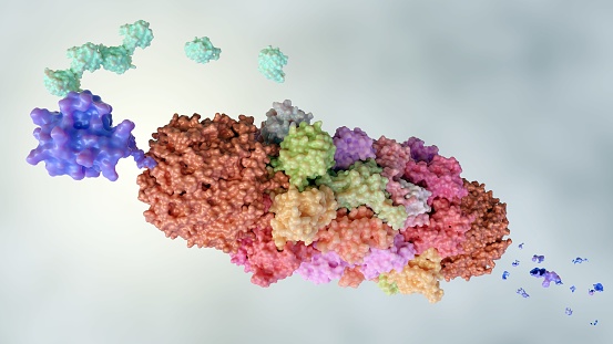
Free Images: "bestof:PDB 2gqv EBI.jpg All beta proteins SH3-like barrel Electron transport accessory proteins R67 dihydrofolate reductase R67 dihydrofolate reductase EBI License"
Methotrexate 1u72.svg
Dihydrofolate reductase reaction.svg
DHFR rxn.svg
Trimetrexate.svg
Aditoprim.svg
Pyrimethamine.svg
Aminopterin.svg
Brodimoprim.svg
Iclaprim.svg
Complex II.svg
Complex II pl.svg
Tetroxoprim.svg
2ph1 pdb gallery nucleotide binding-protein-2 (MinD homolog).svg
P450cycle.svg
PDB 2gqv EBI.jpg
PDB 1vif EBI.jpg
PDB 1vie EBI.jpg
PDB 2rh2 EBI.png
PDB 2rk1 EBI.png
PDB 2rk2 EBI.png
PDB 1dj7 EBI.jpg
PDB 2pu9 EBI.png
PDB 2puo EBI.png
PDB 2puk EBI.png
PDB 2pvo EBI.png
PDB 1psf EBI.jpg
PDB 1gxi EBI.jpg
PDB 1qp3 EBI.jpg
PDB 1qp2 EBI.jpg
PDB 2cz1 EBI.jpg
PDB 2cz6 EBI.jpg
PDB 2cyz EBI.jpg
PDB 2cz0 EBI.jpg
PDB 2d0q EBI.jpg
PDB 2cz7 EBI.jpg
PDB 2ahj EBI.jpg
PDB 1ahj EBI.jpg
PDB 1ugp EBI.jpg
PDB 1ire EBI.jpg
PDB 1ugr EBI.jpg
PDB 1ugs EBI.jpg
PDB 1ugq EBI.jpg
PDB 2zpb EBI.png
PDB 2zpg EBI.png
PDB 2zcf EBI.png
PDB 2zpi EBI.png
PDB 2zpe EBI.png
PDB 2zpf EBI.png
PDB 2zph EBI.png
PDB 1v29 EBI.jpg
PDB 2qdy EBI.png
PDB 1jb0 EBI.jpg
PDB 1ra9 EBI.jpg
PDB 4dfr EBI.jpg
PDB 1ra8 EBI.jpg
PDB 1dra EBI.jpg
PDB 1drb EBI.jpg
PDB 1dhj EBI.jpg
PDB 1rf7 EBI.jpg
PDB 1dyj EBI.jpg
PDB 1dyi EBI.jpg
PDB 1jom EBI.jpg
PDB 1ra1 EBI.jpg
PDB 3drc EBI.jpg
PDB 1dhi EBI.jpg
PDB 1dyh EBI.jpg
PDB 2drc EBI.jpg
PDB 1jol EBI.jpg
PDB 1ra2 EBI.jpg
PDB 1rg7 EBI.jpg
PDB 1ddr EBI.jpg
PDB 1ra3 EBI.jpg
PDB 1dds EBI.jpg
PDB 1rx2 EBI.jpg
PDB 1tdr EBI.jpg
PDB 1rx5 EBI.jpg
PDB 1rc4 EBI.jpg
PDB 1rx9 EBI.jpg
PDB 1rx7 EBI.jpg
PDB 1rx6 EBI.jpg
PDB 1rx1 EBI.jpg
PDB 1rx3 EBI.jpg
PDB 1rx8 EBI.jpg
PDB 1rd7 EBI.jpg
PDB 5dfr EBI.jpg
PDB 1re7 EBI.jpg
PDB 1rb2 EBI.jpg
PDB 1rx4 EBI.jpg
PDB 1rb3 EBI.jpg
PDB 2anq EBI.jpg
PDB 2inq EBI.jpg
PDB 1drh EBI.jpg
PDB 6dfr EBI.jpg
PDB 1rh3 EBI.jpg
PDB 1dre EBI.jpg
PDB 7dfr EBI.jpg
PDB 2ano EBI.jpg
PDB 3dfr EBI.jpg
PDB 1dis EBI.jpg
PDB 1diu EBI.jpg
Terms of Use   Search of the Day