Click Here for More Images from iStock- 15% off with coupon 15FREEIMAGES 
Bacteria Lactobacillus in human intestine,Beneficial healthy intestinal bacterium microflora,Probiotic bacterium
A quadrant streak of the bacterium Staphylococcus aureus cultured or inoculated on Baird-Parker agar with egg tellurite .
Bacterial multidrug efflux pumps (E-CmeB) expel antibiotics  from inside the bacterium (bottom): ciprofloxacin (yellow), chloramphenicol (violet),  ampicillin (red).
Abundant meningococci bacteria observed, with a notably absence of white blood cells in the spinal fluid from a critically ill meningitis patient. Meningococcal meningitis is the term used to describe a bacterial form of meningitis caused by Neisseria meningitidis. This form of meningitis is associated with high morbidity and mortality. \nSometimes the bacteria spread to people who have had close or lengthy contact with a patient with meningococcal disease. Those at increased risk of getting sick include: People in the same household, roommates and anyone with direct contact with the patient’s oral secretions, such as a kissing partner
Branching budding yeast cells with pseudohyphae in  gram stain fine with microscope.
Photomicrograph of filamentous blue-green algae, Oscillatoria species. Each band along the stand is one cell. This filament is very short probably having recently parted from a longer one. Filaments are typically many times longer. This algae is mobile, it moves and twists through the water. San Francisco Bay, California, USA. Live specimen. Wet mount, 40X objective, transmitted brightfield illumination. Note - motion blur of live specimen, very shallow depth of field, chromatic aberration and uneven focus are inherent in light microscopy.
Backgrounds of Bacteria and Mold under the microscope in Lab.
Phospholipid bilayer membrane structures that make up living cells and their contents. This is an image of membrane proteins in the membrane.\nThis image, which looks like a plastic model, evokes biology and biochemistry. It can be used as a cover for magazines, textbooks and presentation materials.\nThis image was not created by AI.
3D cartoon and Gaussian surface models, PDB 6gu2, white background
Cell Membrane Phospholipid Structure,Phospholipid bilayer of the cell membrane,Lipid bilayer,lipid bi-layer cell anatomy
After dopamine binding, the dopamine receptor changes his shape and binds the inactive G protein.The G protein expels its GDP molecule replacing it by GTP. and falls into two pieces. The Galpha subunit binds to and activates the enzyme adenylyl cyclase, which produces then cyclic AMP spreading a signal through the cell.
Close-up shot of a petri dish for checking an antibiotic in the laboratory.
Algae, a unicellular organism
Endosomes - 3d rendered image of Endosomes are membrane-bound organelles. They are parts of endocytic membrane transport pathway.  Endomembrane system in cells. Bilayer Lipid Membrane. Abstract medical illustration. Microscopic view. Microbiologic research concept.
Lily leaf c.s. under microscope
Focal nodular hyperplasia (FNH) is a benign liver lesion that is composed of a proliferation of hyperplastic hepatocytes surrounding a central stellate scar. Typically, FNH is a solitary lesion that is more commonly seen in women.
A quadrant streak of the bacterium Staphylococcus aureus cultured or inoculated on Baird-Parker agar with egg tellurite .
Photomicrograph of rotifer. Rapidly rotating cilia at top of head, internal organs visible. Live specimen. Wet mount, 40X objective, transmitted brightfield illumination.
The Raf/MEK/ERK (MAPK) propagates growth signals in the cell. Misregulation can lead to cancer.\nThe red dots on the active EGFR are phosphorylated tyrosines. PDB entries: 1egf, 1nql, 1m17, 2jwa, 3njp, 2gs6, 1gri, 1xd2, 3ksy, 5p21, 6xi7, 6q0j, 2y4i,
3D cartoon and Gaussian surface models, chain id color scheme, PDB 7t3a, white background
Yeasts sprout in microscopy
Aerial view of loyster farm by Ardara, County Donegal - Ireland.
Close-up shot of a petri dish for checking an antibiotic in the laboratory.
Slime molds, as a group, are polyphyletic under the microscope for education.
Particles, medical concept
From left to right: the NMDA and  AMPA receptors transport calcium cations into neurons after being activated by the neurotransmitter glutamate and  the GABA receptor (right) transport chloride anions after the activation by gamma aminobutyrate. PDB entries: 6wht,  3kg2, 6d6u
Pleomorphic liposarcoma. Site: Neck. Pleomorphic liposarcoma is a rare subtype of fat cell cancer in the body. It is a rare and very malignant type of tumor that usually arises from fat cells and deep soft tissues of the upper and lower extremities (shoulder, arms, legs, and thighs).
Intracellular transport, kinesin motor proteins, orange, transport molecules moving across microtubules, 3D illustration
plant Soya Stem C.S. under light mircoscope with white background
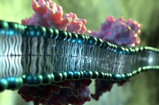
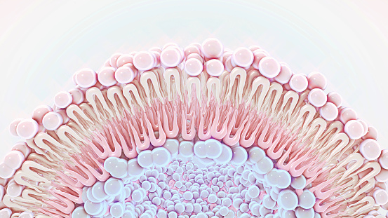
Free Images: "bestof:PDB 2boz EBI.jpg Membrane and cell surface proteins and peptides Bacterial photosystem II reaction centre L and M subunits Bacterial photosystem II reaction"
P450cycle.svg
Luciferin bacterial reaction.svg
Togni s reagent II reaction 01.svg
Togni s reagent II reaction 02.svg
Togni s reagent II reaction 03.svg
Reaction of Propan-1-ol and Copper(II) oxide to Propionaldehyde.svg
Reaction of Propan-1-ol and Copper(II) oxide to Propionaldehyde 2.svg
Fotoukład 2.svg
FMH2-Bioluninescence.svg
PS-Hemmer.svg
Norrish typeII reaction.svg
Ferrier carbocyclization.svg
Alpha oxidation part II.svg
Prokaryote cell diagram international.svg
Bischler-Napieralski Isoquinoline synthesis mechanism II.svg
Semnalizare engl.svg
Hepatitis B virus v2.svg
Taste bud cell types.svg
Deepwater Horizon oil spill recovery context numbered.svg
PDB 2boz EBI.jpg
PDB 2gnu EBI.jpg
PDB 2hg9 EBI.jpg
PDB 2hhk EBI.jpg
PDB 2bns EBI.jpg
PDB 2bnp EBI.jpg
PDB 2hit EBI.jpg
PDB 2gmr EBI.jpg
PDB 2hh1 EBI.jpg
PDB 2hg3 EBI.jpg
PDB 2hj6 EBI.jpg
PDB 1z9k EBI.jpg
PDB 3d38 EBI.png
PDB 1rzh EBI.jpg
PDB 1qov EBI.jpg
PDB 1ry5 EBI.jpg
PDB 1e6d EBI.jpg
PDB 1rg5 EBI.jpg
PDB 1aij EBI.jpg
PDB 1ogv EBI.jpg
PDB 1rqk EBI.jpg
PDB 1kby EBI.jpg
PDB 1rzz EBI.jpg
PDB 1mps EBI.jpg
PDB 1dv3 EBI.jpg
PDB 1ds8 EBI.jpg
PDB 1dv6 EBI.jpg
PDB 1fnq EBI.jpg
PDB 1rgn EBI.jpg
PDB 1fnp EBI.jpg
PDB 1aig EBI.jpg
PDB 1pcr EBI.jpg
PDB 1s00 EBI.jpg
PDB 1jgz EBI.jpg
PDB 1rvj EBI.jpg
PDB 1e14 EBI.jpg
PDB 1jgy EBI.jpg
PDB 1jgw EBI.jpg
PDB 1umx EBI.jpg
PDB 1k6l EBI.jpg
PDB 1f6n EBI.jpg
PDB 1k6n EBI.jpg
PDB 1jgx EBI.jpg
PDB 1m3x EBI.jpg
PDB 1pst EBI.jpg
PDB 1pss EBI.jpg
PDB 1jh0 EBI.jpg
PDB 2rcr EBI.jpg
PDB 1yst EBI.jpg
PDB 4rcr EBI.jpg
PDB 1l9b EBI.jpg
PDB 1l9j EBI.jpg
PDB 2i5n EBI.jpg
PDB 1dxr EBI.jpg
PDB 6prc EBI.jpg
PDB 3prc EBI.jpg
PDB 1prc EBI.jpg
PDB 5prc EBI.jpg
PDB 2prc EBI.jpg
PDB 7prc EBI.jpg
PDB 1r2c EBI.jpg
PDB 1eys EBI.jpg
PDB 1jb0 EBI.jpg
PDB 2qpe EBI.png
PDB 3bvd EBI.png
PDB 2qpd EBI.png
PDB 1m56 EBI.jpg
PDB 1m57 EBI.jpg
PDB 1xme EBI.jpg
PDB 1ehk EBI.jpg
PDB 3dtu EBI.png
PDB 2gsm EBI.jpg
PDB 3ehb EBI.png
PDB 1h2s EBI.jpg
PDB 1h68 EBI.jpg
PDB 2f93 EBI.jpg
PDB 2f95 EBI.jpg
PDB 1gu8 EBI.jpg
PDB 1gue EBI.jpg
PDB 1jgj EBI.jpg
PDB 1qle EBI.jpg
Terms of Use   Search of the Day