Click Here for More Images from iStock- 15% off with coupon 15FREEIMAGES 
GABA B receptors  are G protein-coupled receptors, also called metabotropic receptors.  Binding of  an agonist (baclofen, red) leads to a G-protein coupled C-AMP signaling pathway. Source: PDB entries 7eb2, 6r3q,.
Phospholipid bilayer membrane structures that make up living cells and their contents. This is an image of membrane proteins in the membrane.\nThis image, which looks like a plastic model, evokes biology and biochemistry. It can be used as a cover for magazines, textbooks and presentation materials.\nThis image was not created by AI.
3d helix chromosome or Dna structure, technology science background. 3d render illustration
Chain of Amino Acid. Proteins consist of one or more chains of amino acids called polypeptides
3D cartoon and Gaussian surface models, chain id color scheme, PDB 7t3a, white background
A ball-and-stick plus ribbons molecular model of the hormone Orexin, also known as Hypocretin.  Orexin is involved in the regulation of the sleep/wake cycle and it stimulates apetite, wakefulness and energy expenditure. It has been linked with Alzheimer's disease.  Isolated on white.
Endosomes - 3d rendered image of Endosomes are membrane-bound organelles. They are parts of endocytic membrane transport pathway.  Endomembrane system in cells. Bilayer Lipid Membrane. Abstract medical illustration. Microscopic view. Microbiologic research concept.
Chain of amino acid or bio molecules called protein - 3d illustration
The glucagon like peptide-1 receptor (GLPr) is a G proteincoupled receptor (GPCR) that mediates the action of GLP-1, a peptide hormone. The activated receptor  has a strong effect  on the management of type 2 diabetes mellitus and obesity, including glucose homeostasis and regulation of gastric motility and food intake.
Cell Membrane Phospholipid Structure,Phospholipid bilayer of the cell membrane,Lipid bilayer
Spike protein of SARS-CoV-2, Mutation in the spike protein that the virus uses to bind to the human ACE2 receptor. Mutation sped up spread of coronavirus.
CRISPR-Cas9 gene editing technology
3D cartoon and Gaussian surface models, PDB 5fwk, white background
3d rendering of transparent molecule shape on green color background.
Intracellular transport, kinesin motor proteins, orange, transport molecules moving across microtubules, 3D illustration
ATP Synthase Protein - 3d rendered image. Medical microbiology concept.  Structure and conformational states of the bovine mitochondrial ATP (Adenosine triphosphate) synthase. Chemical energy is synthesized in eukaryotic cells primarily. Hydrolase. Hologram view. HUD, FUI
From left to right: the NMDA and  AMPA receptors transport calcium cations into neurons after being activated by the neurotransmitter glutamate and  the GABA receptor (right) transport chloride anions after the activation by gamma aminobutyrate. PDB entries: 6wht,  3kg2, 6d6u
Focal nodular hyperplasia (FNH) is a benign liver lesion that is composed of a proliferation of hyperplastic hepatocytes surrounding a central stellate scar. Typically, FNH is a solitary lesion that is more commonly seen in women.
Science background with molecule or atom
Photomicrograph of filamentous blue-green algae, Oscillatoria species. Each band along the stand is one cell. This filament is very short probably having recently parted from a longer one. Filaments are typically many times longer. This algae is mobile, it moves and twists through the water. San Francisco Bay, California, USA. Live specimen. Wet mount, 40X objective, transmitted brightfield illumination. Note - motion blur of live specimen, very shallow depth of field, chromatic aberration and uneven focus are inherent in light microscopy.
3D cartoon and Gaussian surface models, PDB 7n94, chain id color scheme, white background
DNA A-T Pair
Dna structure on science background. 3d illustration
A conceptual image of an abstract network of multi-coloured organic strands arranged in a non regular pattern all connected together in a complex manner. The network is made from smaller multi-coloured squares and polyhedra, in vivid colours against a pastel toned background. Shows background and foreground elements to the structure.
The glucagon like peptide-1 receptor (GLPr) is a G proteincoupled receptor (GPCR) that mediates the action of GLP-1, a peptide hormone. The activated receptor  has a strong effect  on the management of type 2 diabetes mellitus and obesity, including glucose homeostasis and regulation of gastric motility and food intake.
DNA Helix - 3d rendered image. Genetic research concept. Complex spiral structure illustration. Medical, science, genetic biotechnology, gene cell, chromosome idea. DNA strands on dark background . Science nanotechnology.
Molecular model of insulin hormone, 3D illustration. It is produced by pancreas and takes part in glucose and lipid metabolism
Phospholipid bilayer membrane structures that make up living cells and their contents. This is an image of membrane proteins in the membrane.\nThis image, which looks like a plastic model, evokes biology and biochemistry. It can be used as a cover for magazines, textbooks and presentation materials.\nThis image was not created by AI.
Spike glycoprotein homotrimer, a main target for neutralization antibody, to bind its receptor, and mediate membrane fusion and virus entry. Surface model, white background.
3D Render of right-handed DNA molecular structures against a white background
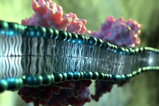
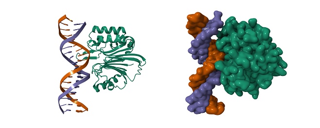
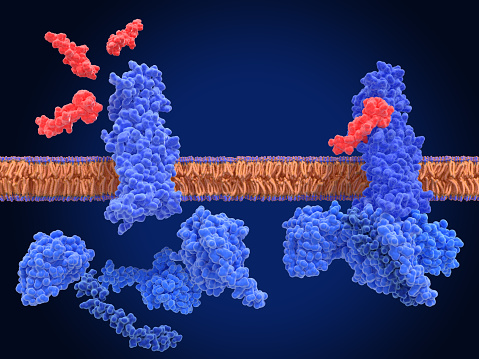
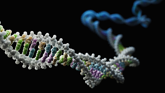
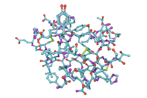
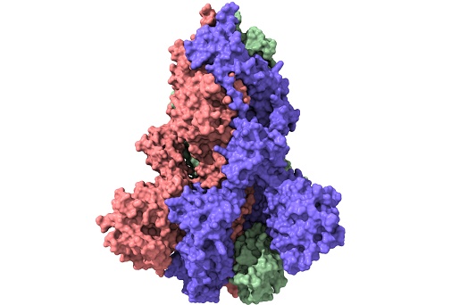
Free Images: "bestof:PDB 2bbj EBI.jpg Membrane and cell surface proteins and peptides Transmembrane helix hairpin Magnesium transport protein CorA transmembrane region Magnesium"
Lipid raft organisation scheme.svg
Human genome by functions.svg
Electron transport chain.svg
Scheme facilitated diffusion in cell membrane-en.svg
Scheme facilitated diffusion in cell membrane-zh.svg
ETC electron transport chain.svg
Scheme facilitated diffusion in cell membrane-eo.svg
2ph1 pdb gallery nucleotide binding-protein-2 (MinD homolog).svg
Scheme facilitated diffusion in cell membrane-zh (zh-tw).svg
Endocytosis types.svg
Exocytosis types.svg
Cell membrane detailed diagram blank.svg
Intermediate filament.svg
Scheme facilitated diffusion in cell membrane-ca.svg
Hepatitis B virus v2.svg
Insulin glucose metabolism ZP.svg
Cellular tight junction keys.svg
Insulin glucose metabolism ZP-ca.svg
PDB 2bbj EBI.jpg
PDB 2iub EBI.jpg
PDB 2hn2 EBI.jpg
PDB 2qpe EBI.png
PDB 3bvd EBI.png
PDB 2qpd EBI.png
PDB 1jb0 EBI.jpg
PDB 3dtu EBI.png
PDB 3ehb EBI.png
PDB 3cx5 EBI.png
PDB 3cxh EBI.png
PDB 1a91 EBI.jpg
PDB 1ijp EBI.jpg
PDB 1c99 EBI.jpg
PDB 1c17 EBI.jpg
PDB 1l6t EBI.jpg
PDB 1c0v EBI.jpg
PDB 1ppj EBI.jpg
PDB 2a06 EBI.jpg
PDB 1t16 EBI.jpg
PDB 1t1l EBI.jpg
PDB 2hyd EBI.jpg
PDB 2onj EBI.jpg
PDB 1rzh EBI.jpg
PDB 1qov EBI.jpg
PDB 1ry5 EBI.jpg
PDB 1e6d EBI.jpg
PDB 1rg5 EBI.jpg
PDB 1aij EBI.jpg
PDB 1ogv EBI.jpg
PDB 1rqk EBI.jpg
PDB 1kby EBI.jpg
PDB 1rzz EBI.jpg
PDB 1mps EBI.jpg
PDB 1dv3 EBI.jpg
PDB 1ds8 EBI.jpg
PDB 1dv6 EBI.jpg
PDB 1fnq EBI.jpg
PDB 1rgn EBI.jpg
PDB 1fnp EBI.jpg
PDB 1aig EBI.jpg
PDB 1pcr EBI.jpg
PDB 1s00 EBI.jpg
PDB 1jgz EBI.jpg
PDB 1rvj EBI.jpg
PDB 1e14 EBI.jpg
PDB 1jgy EBI.jpg
PDB 1jgw EBI.jpg
PDB 1umx EBI.jpg
PDB 1k6l EBI.jpg
PDB 1f6n EBI.jpg
PDB 1k6n EBI.jpg
PDB 1jgx EBI.jpg
PDB 1m3x EBI.jpg
PDB 1pst EBI.jpg
PDB 1pss EBI.jpg
PDB 1jh0 EBI.jpg
PDB 2rcr EBI.jpg
PDB 1yst EBI.jpg
PDB 4rcr EBI.jpg
PDB 2e74 EBI.jpg
PDB 2e76 EBI.jpg
PDB 2e75 EBI.jpg
PDB 2d2c EBI.jpg
PDB 1l9b EBI.jpg
PDB 1l9j EBI.jpg
PDB 3d38 EBI.png
PDB 1pp9 EBI.jpg
PDB 1ntm EBI.jpg
PDB 1l0l EBI.jpg
PDB 1ntz EBI.jpg
PDB 1ntk EBI.jpg
PDB 1sqb EBI.jpg
PDB 1l0n EBI.jpg
PDB 1nu1 EBI.jpg
PDB 1be3 EBI.jpg
PDB 1bgy EBI.jpg
PDB 1qcr EBI.jpg
PDB 1bcc EBI.jpg
PDB 2bcc EBI.jpg
PDB 3bcc EBI.jpg
PDB 2oau EBI.jpg
Terms of Use   Search of the Day