Click Here for More Images from iStock- 15% off with coupon 15FREEIMAGES 
Chain of Amino Acid. Proteins consist of one or more chains of amino acids called polypeptides
3d helix chromosome or Dna structure, technology science background. 3d render illustration
3D cartoon and Gaussian surface models, chain id color scheme, PDB 7t3a, white background
Phospholipid bilayer membrane structures that make up living cells and their contents. This is an image of membrane proteins in the membrane.\nThis image, which looks like a plastic model, evokes biology and biochemistry. It can be used as a cover for magazines, textbooks and presentation materials.\nThis image was not created by AI.
From left to right: the NMDA and  AMPA receptors transport calcium cations into neurons after being activated by the neurotransmitter glutamate and  the GABA receptor (right) transport chloride anions after the activation by gamma aminobutyrate. PDB entries: 6wht,  3kg2, 6d6u
A ball-and-stick plus ribbons molecular model of the hormone Orexin, also known as Hypocretin.  Orexin is involved in the regulation of the sleep/wake cycle and it stimulates apetite, wakefulness and energy expenditure. It has been linked with Alzheimer's disease.  Isolated on white.
Endosomes - 3d rendered image of Endosomes are membrane-bound organelles. They are parts of endocytic membrane transport pathway.  Endomembrane system in cells. Bilayer Lipid Membrane. Abstract medical illustration. Microscopic view. Microbiologic research concept.
Intracellular transport, kinesin motor proteins, orange, transport molecules moving across microtubules, 3D illustration
Dna structure on science background. 3d illustration
3d rendering of transparent molecule shape on green color background.
Proliferating cell nuclear antigen (PCNA) is a DNA clamp that acts as a processivity factor for DNA polymerase δ in eukaryotic cells and is essential for replication. PCNA is a homotrimer and achieves its processivity by encircling the DNA, where it acts as a scaffold to recruit proteins involved in DNA replication, DNA repair, chromatin remodeling and epigenetics. 3D cartoon and Gaussian surface models, chain instance color scheme, based on PDB 3wgw, white background.
Cell Membrane Phospholipid Structure,Phospholipid bilayer of the cell membrane,Lipid bilayer
After dopamine binding, the dopamine receptor changes his shape and binds the inactive G protein.The G protein expels its GDP molecule replacing it by GTP. and falls into two pieces. The Galpha subunit binds to and activates the enzyme adenylyl cyclase, which produces then cyclic AMP spreading a signal through the cell.
CRISPR-Cas9 gene editing technology
Ball and stick 3D render of the caffeine molecular structure.
Phospholipid bilayer membrane structures that make up living cells and their contents. This is an image of membrane proteins in the membrane.\nThis image, which looks like a plastic model, evokes biology and biochemistry. It can be used as a cover for magazines, textbooks and presentation materials.\nThis image was not created by AI.
Spirogyra sp. Algae under microscopic view x40, Chlorophyta, green algae, blue green algae, background
Futuristic artificial DNA strand among organic models. Medical innovation and technology concept.
3D cartoon and Gaussian surface models, PDB 7n94, chain id color scheme, white background
Illustration of DNA.
The glucagon like peptide-1 receptor (GLPr) is a G proteincoupled receptor (GPCR) that mediates the action of GLP-1, a peptide hormone. The activated receptor  has a strong effect  on the management of type 2 diabetes mellitus and obesity, including glucose homeostasis and regulation of gastric motility and food intake.
DNA strands magnified.
Science background with molecule or atom
Dna structure digital concept
Nucleosome structure. Structure of an oligonucleosome, showing the packaging of DNA in chromosomes. 3D illustration.
ATP Synthase Protein - 3d rendered image. Medical microbiology concept.  Structure and conformational states of the bovine mitochondrial ATP (Adenosine triphosphate) synthase. Chemical energy is synthesized in eukaryotic cells primarily. Hydrolase. Hologram view. HUD, FUI
Spike glycoprotein homotrimer, a main target for neutralization antibody, to bind its receptor, ACE2, and mediate membrane fusion and virus entry. 3D cartoon and Gaussian surface models, white background.
DNA and biotechnology concept
The calcitonin gene related peptide (yellow) binds to its receptor (blue) on neurons and smooth muscle cells of cerebral blood vessels, activating a signal cascade through G-proteins (dark blue) in this cells that  leads to a dilatation of the blood vessels (vasodilatation).
DNA Molecule: a 3D illustration of the double helix structure reflecting the genetic code. This 3D illustration is a visualization of the DNA molecule showing its characteristic double helix structure. The model reflects the major components of DNA such as the nitrogenous bases (adenine, thymine, guanine, and cytosine), sugars, and phosphate groups that together form the genetic code. This illustration can be used for educational purposes, scientific publications or medical advertising to demonstrate the complexity and beauty of the molecule responsible for heredity and the development of life on Earth.
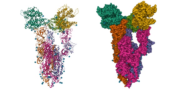
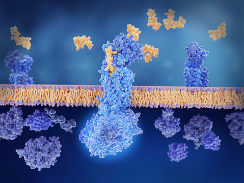
Free Images: "bestof:PDB 1ppj EBI.jpg Membrane and cell surface proteins and peptides Single transmembrane helix Cytochrome c1 subunit of cytochrome bc1 complex Ubiquinol-cytochrome"
Electron transport chain.svg
ETC electron transport chain.svg
P450cycle.svg
Lipid raft organisation scheme.svg
Hepatitis B virus v2.svg
Cellular tight junction keys.svg
PDB 1ppj EBI.jpg
PDB 2a06 EBI.jpg
PDB 2fyu EBI.jpg
PDB 1sqx EBI.jpg
PDB 1sqv EBI.jpg
PDB 1sqp EBI.jpg
PDB 1sqq EBI.jpg
PDB 1p84 EBI.jpg
PDB 1kb9 EBI.jpg
PDB 2ibz EBI.jpg
PDB 1ezv EBI.jpg
PDB 1kyo EBI.jpg
PDB 1pp9 EBI.jpg
PDB 1ntm EBI.jpg
PDB 1l0l EBI.jpg
PDB 1ntz EBI.jpg
PDB 1ntk EBI.jpg
PDB 1sqb EBI.jpg
PDB 1l0n EBI.jpg
PDB 1nu1 EBI.jpg
PDB 1be3 EBI.jpg
PDB 1bgy EBI.jpg
PDB 1qcr EBI.jpg
PDB 1bcc EBI.jpg
PDB 2bcc EBI.jpg
PDB 3bcc EBI.jpg
PDB 3cx5 EBI.png
PDB 3cxh EBI.png
PDB 1vf5 EBI.jpg
PDB 1q90 EBI.jpg
PDB 2e74 EBI.jpg
PDB 2e76 EBI.jpg
PDB 2e75 EBI.jpg
PDB 2d2c EBI.jpg
PDB 2qpe EBI.png
PDB 3bvd EBI.png
PDB 2qpd EBI.png
PDB 1m56 EBI.jpg
PDB 1m57 EBI.jpg
PDB 1xme EBI.jpg
PDB 1ehk EBI.jpg
PDB 1qle EBI.jpg
PDB 1l9b EBI.jpg
PDB 1l9j EBI.jpg
PDB 1v54 EBI.jpg
PDB 2dyr EBI.jpg
PDB 1v55 EBI.jpg
PDB 2eij EBI.jpg
PDB 2eil EBI.jpg
PDB 2eik EBI.jpg
PDB 2dys EBI.jpg
PDB 2occ EBI.jpg
PDB 1ocr EBI.jpg
PDB 2eim EBI.jpg
PDB 1occ EBI.jpg
PDB 2ein EBI.jpg
PDB 1ocz EBI.jpg
PDB 1oco EBI.jpg
PDB 2i5n EBI.jpg
PDB 1dxr EBI.jpg
PDB 6prc EBI.jpg
PDB 3prc EBI.jpg
PDB 1prc EBI.jpg
PDB 5prc EBI.jpg
PDB 2prc EBI.jpg
PDB 7prc EBI.jpg
PDB 1r2c EBI.jpg
PDB 1eys EBI.jpg
PDB 3d38 EBI.png
PDB 2bs2 EBI.jpg
PDB 1qlb EBI.jpg
PDB 1e7p EBI.jpg
PDB 3dtu EBI.png
PDB 3ehb EBI.png
PDB 1rkl EBI.jpg
PDB 1l2p EBI.jpg
PDB 1rh5 EBI.jpg
PDB 1rhz EBI.jpg
PDB 1kqf EBI.jpg
PDB 1kqg EBI.jpg
PDB 1rzh EBI.jpg
PDB 1qov EBI.jpg
PDB 1ry5 EBI.jpg
PDB 1e6d EBI.jpg
PDB 1rg5 EBI.jpg
PDB 1aij EBI.jpg
PDB 1ogv EBI.jpg
PDB 1rqk EBI.jpg
PDB 1kby EBI.jpg
PDB 1rzz EBI.jpg
PDB 1mps EBI.jpg
PDB 1dv3 EBI.jpg
PDB 1ds8 EBI.jpg
PDB 1dv6 EBI.jpg
Terms of Use   Search of the Day