Click Here for More Images from iStock- 15% off with coupon 15FREEIMAGES 
The LDL-receptor (red) is a membrane protein found in almost every human cell. It binds to the apoprotein B100 from LDL-particles and mediates their internalization (endocytosis) in the cell. This occurs in almost all cells but mainly in the liver where it removes about 70 % of LDL from the circulation.
The three-dimensional structure of proteins is represented in a variety of conformations. This image is based on the ribbon structure of a protein. It is suitable for biology notebooks and book covers.\nThis image has not been created by AI.
Proteasomes are molecular machines for breaking down proteins called proteolysis, only target proteins that have been marked for destruction called ubiquitin to the target protein, 3d rendering
Models of an antibody broadly antagonistic to HIV.  A challenge in AIDS and HIV research is producing a vaccine which elicits antibodies effective against the many variations of HIV proteins.Isolated on white.
3D cartoon and Gaussian surface models, chain id color scheme, PDB 7t3a, white background
Photomicrograph of filamentous blue-green algae, Oscillatoria species. Each band along the stand is one cell. This filament is very short probably having recently parted from a longer one. Filaments are typically many times longer. This algae is mobile, it moves and twists through the water. San Francisco Bay, California, USA. Live specimen. Wet mount, 40X objective, transmitted brightfield illumination. Note - motion blur of live specimen, very shallow depth of field, chromatic aberration and uneven focus are inherent in light microscopy.
Endosomes - 3d rendered image of Endosomes are membrane-bound organelles. They are parts of endocytic membrane transport pathway.  Endomembrane system in cells. Bilayer Lipid Membrane. Abstract medical illustration. Microscopic view. Microbiologic research concept.
3d rendered Gastric inhibitory polypeptide (GIP, glucose-dependent insulinotropic peptide) endocrine protein hormone, 3D illustration
orange: RNA primer; red: RNA template; light blue: proofreading riboexonuclease; mid blue: helicase; dark blue: RNA directed RNA polymerase; violet: non structural proteins (NSPs).
Phospholipid bilayer membrane structures that make up living cells and their contents. This is an image of membrane proteins in the membrane.\nThis image, which looks like a plastic model, evokes biology and biochemistry. It can be used as a cover for magazines, textbooks and presentation materials.\nThis image was not created by AI.
DNA close-up, DNA Molecule Model, DNA Strands, DNA animation
Focal nodular hyperplasia (FNH) is a benign liver lesion that is composed of a proliferation of hyperplastic hepatocytes surrounding a central stellate scar. Typically, FNH is a solitary lesion that is more commonly seen in women.
Ferritin is a protein that contains iron and is the primary form of iron stored inside of cells. 3D cartoon model, PDB 4y08, white background
Covid-19 Coronavirus Variants Omicron Delta Gamma Beta Alpha Pandemic Virus Disease Mutation
IG-g like Antibodies are attacking a foreign protein.
Endosomes - 3d rendered image of Endosomes are membrane-bound organelles. They are parts of endocytic membrane transport pathway.  Endomembrane system in cells. Bilayer Lipid Membrane. Abstract medical illustration. Microscopic view. Microbiologic research concept.
GABA B receptors  are G protein-coupled receptors, also called metabotropic receptors.  Binding of  an agonist (baclofen, red) leads to a G-protein coupled C-AMP signaling pathway. Source: PDB entries 7eb2, 6r3q,.
Particles, medical concept
Nucleosome structure. Structure of an oligonucleosome, showing the packaging of DNA in chromosomes. 3D illustration.
Digital background depicting innovative technologies in (AI) artificial systems, neural interfaces and internet machine learning technologies
3D cartoon and Gaussian surface models, PDB 6vg8, white background
Photomicrograph of rotifer. Rapidly rotating cilia at top of head, internal organs visible. Live specimen. Wet mount, 40X objective, transmitted brightfield illumination.
Dna structure on science background. 3d illustration
Abs Virus - 3d rendered image like colorized scanning electron micrograph. Scene showing virus (yellow)  emerging from infected cell.\nConcept of coronavirus that’s causing a global pandemic. Covid-19, Sars-cov-2 alpha, betta, gamma, delta, lambda variant, 2019-nCoV, Coronavirus.\nSEM, TEM, hologram view.
Bacterial multidrug efflux pumps (E-CmeB) expel antibiotics  from inside the bacterium (bottom): ciprofloxacin (yellow), chloramphenicol (violet),  ampicillin (red).
anaplastic thyroid carcinoma with heterologous differentiation (leiomyosarcoma)
DNA close-up, DNA Molecule Model, DNA Strands, DNA animation
Cells in the body examined by endoscope
3D cartoon and Gaussian surface models, chain id color scheme, PDB 3omv, white background
Liver Fluke Section under light microscope with white background
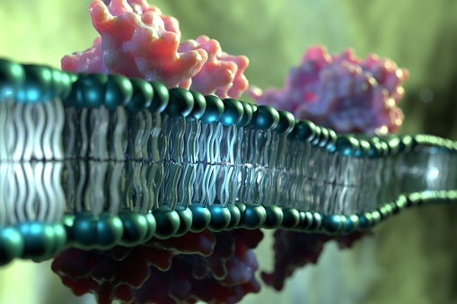
Free Images: "bestof:PDB 1lv3 EBI.jpg Small proteins Glucocorticoid receptor-like DNA-binding domain Glucocorticoid receptor-like DNA-binding domain Hypothetical zinc finger protein"
Notable mutations.svg
Hepatitis B virus v2.svg
Trifluoroisopropanolamine SEGRA.svg
MK-5932 skeletal.svg
A 276575 structure.svg
ZK 216348 structure.svg
RU 24858 skeletal.svg
Octahydrophenanthrene-2,7-diol SEGRA.svg
Mapracorat skeletal.svg
Stat domain structure (zh-cn).svg
Insulin glucose metabolism ZP.svg
Insulin glucose metabolism ZP-ca.svg
Complement pathway.svg
PDB 1lv3 EBI.jpg
PDB 1tjl EBI.jpg
PDB 2das EBI.png
PDB 1pzw EBI.jpg
PDB 1uw0 EBI.jpg
PDB 1xbt EBI.jpg
PDB 2orv EBI.jpg
PDB 2b8t EBI.jpg
PDB 2uz3 EBI.jpg
PDB 1xx6 EBI.jpg
PDB 2fif EBI.jpg
PDB 2fid EBI.jpg
PDB 2c7n EBI.jpg
PDB 2c7m EBI.jpg
PDB 1r2z EBI.jpg
PDB 1l1t EBI.jpg
PDB 1l2c EBI.jpg
PDB 2f5q EBI.jpg
PDB 2f5s EBI.jpg
PDB 1tdz EBI.jpg
PDB 1xc8 EBI.jpg
PDB 1pm5 EBI.jpg
PDB 1kfv EBI.jpg
PDB 1ee8 EBI.jpg
PDB 1l1z EBI.jpg
PDB 1l2d EBI.jpg
PDB 1r2y EBI.jpg
PDB 1l2b EBI.jpg
PDB 1k82 EBI.jpg
PDB 1pjj EBI.jpg
PDB 1pji EBI.jpg
PDB 1nnj EBI.jpg
PDB 2ds5 EBI.jpg
PDB 2ds8 EBI.jpg
PDB 2ds6 EBI.jpg
PDB 1ovx EBI.jpg
PDB 1ctl EBI.jpg
PDB 1b8t EBI.jpg
PDB 1ibi EBI.jpg
PDB 1cxx EBI.jpg
PDB 1a7i EBI.jpg
PDB 1qli EBI.jpg
PDB 1iml EBI.jpg
PDB 2cu8 EBI.jpg
PDB 2dar EBI.jpg
PDB 1x62 EBI.jpg
PDB 2co8 EBI.jpg
PDB 1x64 EBI.jpg
PDB 2d8r EBI.png
PDB 2d8x EBI.jpg
PDB 2cor EBI.jpg
PDB 1nyp EBI.jpg
PDB 1u5s EBI.jpg
PDB 1g47 EBI.jpg
PDB 1v6g EBI.jpg
PDB 1wig EBI.jpg
PDB 2d8z EBI.jpg
PDB 1x4k EBI.jpg
PDB 1x4l EBI.jpg
PDB 2cur EBI.jpg
PDB 1x63 EBI.jpg
PDB 2cup EBI.jpg
PDB 1x61 EBI.jpg
PDB 2dlo EBI.jpg
PDB 2dj7 EBI.jpg
PDB 1x68 EBI.jpg
PDB 2gvi EBI.png
PDB 2b9m EBI.jpg
PDB 2b9o EBI.jpg
PDB 1n32 EBI.jpg
PDB 1xnr EBI.jpg
PDB 1hnz EBI.jpg
PDB 1hnw EBI.jpg
PDB 1hnx EBI.jpg
PDB 1xmq EBI.jpg
PDB 1n34 EBI.jpg
PDB 1n36 EBI.jpg
PDB 1i95 EBI.jpg
PDB 1j5e EBI.jpg
PDB 1xnq EBI.jpg
PDB 1i94 EBI.jpg
PDB 1xmo EBI.jpg
PDB 1fjg EBI.jpg
PDB 1n33 EBI.jpg
PDB 1i96 EBI.jpg
PDB 1i97 EBI.jpg
PDB 2uuc EBI.jpg
Terms of Use   Search of the Day