Click Here for More Images from iStock- 15% off with coupon 15FREEIMAGES 
The inner layer of the eye, or retina, is similar to film in a camera. It receives light from an image we are looking at, and converts that light into electrical impulses which are sent through the fibers of the optic nerve to the brain.
Using a biomicroscope to evaluate the patient's eyes.
Eye test device on table alongside Snellen chart in office of ophthalmologist. Office of eye doctor provides care to enhance health of patients
Human eye and graphical interface. Smart wearable technology concept
Ophthalmologist examining patient in his office - Buenos Aires - Argentina
Bestrophinopathies, inherited retinal disorders caused by mutations in the BEST1 gene, illustration. Best disease, autosomal recessive bestrophinopathy and autosomal dominant vitreoretinochoroidopathy
Doctor and patient in ophthalmology clinic
Phoropter, hospital and eye exam for health, check and lens for vision, wellness and helping with machine. Ophthalmology, glasses and test with healthcare, metal frame and care for eyesight in clinic
Patient on diagnostic of vision, oculist cabinet. Optician measuring a woman's eyesight. Young woman visiting ophthalmologist in clinic. Optometrist checking vision of young beautiful woman.
Close-up on a pair of eyeglasses on an eye chart - eye exam concepts
High quality raster illustration of retinal detachment with clipping path.
Photo of optometrist examining patient's eyes in ophthalmology clinic.
How images are captured by our eyes and processed by our brain. Digital illustration.
Doctor and patient in ophthalmology clinic
Medical student performing an eye exam on a patient at a teaching hospital - medical school concepts
3D illustration of a cross-section of the human eye in a side view and a frontal view
Phoropter for eye test, lenses glasses for eye check in optical store
Male ophthalmologist examining eyes of a young patient using refractometer during a medical examination in the office.
Retinal scar in toxoplasmosis, a disease caused by the single-celled protozoan Toxoplasma gondii, and the same healthy eye retina for comparison, fluorescein angiography, scientific illustration
Mother with two kids at the Ophthalmologist for an eye check up
Optometrist diopter. optical phoropter
Beautiful young girl checking the eye vision in modern ophthalmology clinic
Patient getting an eye exam at the ophthalmologist - healthcare and medicine concepts
Female patient facing slit lamp while her chin and forehead resting on support during ophthalmic examination done by optometrist
Eye anatomy - inner structure isolated on white
Man having his eye structures examined by a special optometrist equipment
High technology concept health for eyes care - The optician ophthalmology doctor optometrist in the eyes clinic using a binocular slit-lamp to a young woman for an eye control.
Ophthalmologist examining patient's eyes. Side view of White man is checking the eye vision with phoropter eyesight measurement testing machine. Medical check up. Handsome man getting an eye exam at ophthalmology clinic. Checking retina of a male eye close-up.
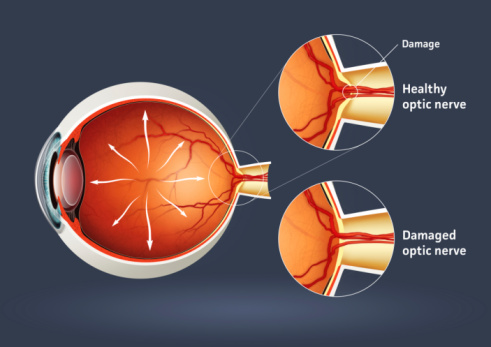
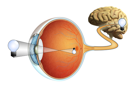
Free Images: "bestof:Owlretina-hu.svg diagram of the retina of an owl - Hungarian version File Owlretina svg 17 48 12 May 2010 UTC User Syp File Owlretina svg Bird visual system"
Terms of Use   Search of the Day