Click Here for More Images from iStock- 15% off with coupon 15FREEIMAGES 
3D computer graphic sagittal section of female reproductive system with labels.
Chronic viral hepatitis B. Chronic hepatitis B infection lasts six months or longer. It lingers because your immune system can't fight off the infection. Chronic hepatitis B infection may last a lifetime, possibly leading to serious illnesses such as cirrhosis and liver cancer.
thyrohyoid membrane connects the thyroid cartilage to the hyoid bone and facilitates the superior movement of the larynx during 3d illustration
plant Zea Stem C.S. under light mircoscope with white background
Conceptual image for viral ethiology of prostate cancer. 3D illustration showing viruses infecting prostate gland which develops cancerous tumor
skin science 3d
Leiden leaf c.s. under microscope
Texture Pattern Design on Banknote
Micrograph of rat brain. Science cross section.
Carcinoid tumors are a type of slow-growing cancer that can arise in several places throughout your body. Carcinoid tumors, which are one subset of tumors called neuroendocrine tumors, usually begin in the digestive tract (stomach, appendix, small intestine, colon, rectum) or in the lungs. Site: Ovary
Cross section leaf of plant under the light microscope view for botany education.
Tetis of Fish T.S. under light microscope with white background
Microscope photo of a sambucus stem with parenchyma cells.
Micro world
Adipose tissue human, Smooth muscles human, Bone human and Striated (skeletal) muscle human under the microscope in Lab.
Photomicrograph of filamentous blue-green algae, Oscillatoria species. Each band along the stand is one cell. This filament is very short probably having recently parted from a longer one. Filaments are typically many times longer. This algae is mobile, it moves and twists through the water. San Francisco Bay, California, USA. Live specimen. Wet mount, 40X objective, transmitted brightfield illumination. Note - motion blur of live specimen, very shallow depth of field, chromatic aberration and uneven focus are inherent in light microscopy.
forming of underskin cellulite illustration
anaplastic thyroid carcinoma with heterologous differentiation (leiomyosarcoma)
Microscopic close view of high vaginal swab Gram stain smear, 100x. diagnosis of Bacterial vaginosis (BV).
plant rape flower cross section under light mircoscope with white background
cross section cut of pig embryo
Clockwise, axial and sagittal T1 post contrast, then sagittal and axial T2 MRI of the pelvis with a large (11 cm) uterine fibroid with central necrosis, compressing the urinary bladder and rectum. Uterine fibroids are common benign tumors of the uterus that first appear during fertile years and grow with hormonal stimulation until menopause. They can be tiny and go unnoticed or grow as large as a full-term baby. In addition to expanding the waistline, fibroids can cause prolonged, heavy menstrual bleeding and pain, pelvic pain, and pressure on the bladder and rectum leading to difficulty with passing bowel movements and frequent urination. They can impair venous blood flow out of the legs, causing leg pain and heaviness. Most fibroids can be treated with a non-invasive procedure where the blood supply is cut off through a catheter threaded from the femoral artery into the uterine artery, termed uterine artery embolization or uterine fibroid embolization, performed by an interventional radiologist.
The detrusor muscle remains relaxed to allow the bladder to store urine, and contracts during urination to release urine 3d illustration
Photomicrograph of two rotifers. Rapidly rotating cilia at top of heads, internal organs visible. Live specimen. Wet mount, 10X objective, transmitted brightfield illumination.
Backgrounds of Characteristics Tissue Tongue, Small intestine of Cat and Tissue of Esophagus rabbit under microscope.
Pheochromocytoma, Site: Adrenal gland
Dirty pore, main trouble of blackhead and acne. Skin treatment, large pores 3D rendering
PAPS Smear study of a young women under microscopy showing Atrophic changes in uterus which is unusual at young age.
flower bud of lilium brownii var. viridulum T.S. on microscopy
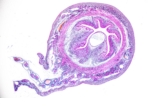
Free Images: "bestof:Origin of Vertebrates Fig 098.png Fig 98 �Transverse Section through the Olfactory Passage of Petromyzon cart nasal cartilage All references in this work to"
Hegi_placentation.svg
Gray506_pl.svg
Wound healing phases.svg
George_Pyne_-_George_James_Drummond's_Room_at_Oxford,_1853_-_Google_Art_Project_(GgH0jYSQxu0IwQ).jpg
İmparator_Süleyman’ın_Kızı_Cameria_(Mihrimah_Sultan).jpg
Rattus_macleari_skull_labeled.svg
Profil_rail_funiculaire.svg
Swine flu infection exponent by county June 2009.svg
Origin_of_Vertebrates_Fig_098.png
Origin_of_Vertebrates_Fig_099.png
Origin_of_Vertebrates_Fig_065.png
Origin_of_Vertebrates_Fig_126.png
Origin_of_Vertebrates_Fig_118.jpg
Origin_of_Vertebrates_Fig_134.jpg
Origin_of_Vertebrates_Fig_056.png
Origin_of_Vertebrates_Fig_131.png
Origin_of_Vertebrates_Fig_022.png
Origin_of_Vertebrates_Fig_141.png
Origin_of_Vertebrates_Fig_154.png
Origin_of_Vertebrates_Fig_116.jpg
Origin_of_Vertebrates_Fig_031.jpg
Origin_of_Vertebrates_Fig_071.jpg
Origin_of_Vertebrates_Fig_124.jpg
Origin_of_Vertebrates_Fig_074.jpg
Origin_of_Vertebrates_Fig_057.jpg
Origin_of_Vertebrates_Fig_055.jpg
Origin_of_Vertebrates_Fig_052.png
Origin_of_Vertebrates_Fig_090.png
Origin_of_Vertebrates_Fig_133.png
Origin_of_Vertebrates_Fig_073.png
Origin_of_Vertebrates_Fig_132.png
Origin_of_Vertebrates_Fig_117.jpg
Origin_of_Vertebrates_Fig_106.png
Origin_of_Vertebrates_Fig_034.png
Origin_of_Vertebrates_Fig_122.png
Origin_of_Vertebrates_Fig_140.png
Origin_of_Vertebrates_Fig_053.png
Origin_of_Vertebrates_Fig_135.png
Origin_of_Vertebrates_Fig_033.png
Origin_of_Vertebrates_Fig_125.png
Origin_of_Vertebrates_Fig_068.png
Origin_of_Vertebrates_Fig_081.png
Origin_of_Vertebrates_Fig_097.png
Origin_of_Vertebrates_Fig_066.png
Origin_of_Vertebrates_Fig_063.jpg
Origin_of_Vertebrates_Fig_018.png
Origin_of_Vertebrates_Fig_025.png
Origin_of_Vertebrates_Fig_037.png
Origin_of_Vertebrates_Fig_054.png
Origin_of_Vertebrates_Fig_092.png
Origin_of_Vertebrates_Fig_119.png
Origin_of_Vertebrates_Fig_121.png
Origin_of_Vertebrates_Fig_060.jpg
Origin_of_Vertebrates_Fig_064.jpg
Origin_of_Vertebrates_Fig_115.jpg
Origin_of_Vertebrates_Fig_123.jpg
Origin_of_Vertebrates_Fig_020.png
Origin_of_Vertebrates_Fig_069.png
Origin_of_Vertebrates_Fig_080.png
Origin_of_Vertebrates_Fig_082.png
Origin_of_Vertebrates_Fig_083.png
Origin_of_Vertebrates_Fig_084.png
Origin_of_Vertebrates_Fig_093.png
Origin_of_Vertebrates_Fig_162.png
Origin_of_Vertebrates_Fig_163.png
Origin_of_Vertebrates_Fig_114.jpg
Origin_of_Vertebrates_Fig_041.png
Origin_of_Vertebrates_Fig_047.png
Origin_of_Vertebrates_Fig_085.png
Origin_of_Vertebrates_Fig_100.png
Origin_of_Vertebrates_Fig_113.png
Origin_of_Vertebrates_Fig_120.png
Origin_of_Vertebrates_Fig_049.png
Origin_of_Vertebrates_Fig_050.png
Origin_of_Vertebrates_Fig_048.png
Origin_of_Vertebrates_Fig_019.png
Origin_of_Vertebrates_Fig_166.png
Origin_of_Vertebrates_Fig_023.png
Origin_of_Vertebrates_Fig_026.png
Origin_of_Vertebrates_Fig_045.png
Origin_of_Vertebrates_Fig_058.png
Origin_of_Vertebrates_Fig_070.png
Origin_of_Vertebrates_Fig_086.png
Origin_of_Vertebrates_Fig_087.png
Origin_of_Vertebrates_Fig_088.png
Origin_of_Vertebrates_Fig_089.png
Origin_of_Vertebrates_Fig_091.png
Origin_of_Vertebrates_Fig_096.png
Origin_of_Vertebrates_Fig_105.png
Origin_of_Vertebrates_Fig_130.png
Origin_of_Vertebrates_Fig_146.png
Origin_of_Vertebrates_Fig_147.png
Origin_of_Vertebrates_Fig_148.png
Origin_of_Vertebrates_Fig_150.png
Origin_of_Vertebrates_Fig_152.png
Origin_of_Vertebrates_Fig_153.png
Origin_of_Vertebrates_Fig_157.png
Origin_of_Vertebrates_Fig_158.png
Origin_of_Vertebrates_Fig_160.png
Origin_of_Vertebrates_Fig_167.png
Terms of Use   Search of the Day