Click Here for More Images from iStock- 15% off with coupon 15FREEIMAGES 
Photographic scan of a human retina. In the middle is the optic nerve, with blood vessels coming out.
Bestrophinopathies, inherited retinal disorders caused by mutations in the BEST1 gene, illustration. Best disease, autosomal recessive bestrophinopathy and autosomal dominant vitreoretinochoroidopathy
Light microscope micrograph of a retina stained with the Golgi method. In the inner layer there are many amacrine cells with pear-shaped cell bodies located in the inner nuclear layer and dendrites ending in the inner plexiform layer.
Blastomycosis is a fungal infection caused by inhaling Blastomyces dermatitidis spores. If it involves only the lungs, it is called pulmonary blastomycosis. Only about half of people with the disease have symptoms, which can include fever, cough, night sweats, muscle pains, weight loss, chest pain, and feeling tired.
Plant tissue Structure, section (tissue) of stem plant tissue under a light microscope.
Photomicrograph of algae, Cosmarium turpinii, a desmid. Live specimen. Wet mount, 40X objective, transmitted brightfield illumination.
plant Zea Stem C.S. under light mircoscope with white background
Colorful, abstract micrograph of a fish scale, calico bass,Paralabrax clathratus, with polarization at 100x.
Freshwater aquatic zooplankton and algae under microscope view
Trematode parasite under a microscope
Optical Coherence Tomography . ( OCT )
anaplastic thyroid carcinoma with heterologous differentiation (leiomyosarcoma)
High quality raster illustration of retinal detachment with clipping path.
Firing Neurons - 3d rendered image of Neuron cell network on black background.  Conceptual medical illustration.  Healthcare concept. \nAbstract stylized npr (non photorealistic rendering) view. Half Tone, Spotted, Stipple Effect. Funky, popart, retro style colors.
Leiden leaf c.s. under microscope
Liver Fluke Section under light microscope with white background
Micrograph of rat brain. Science cross section.
Computational tomography image of the abdominal and pelvic regions.
Epidermis. Electron microscope micrograph showing a keratinocyte of spinous layer. The epithelial cell has a polygonal shape, central nucleus with nucleolus, cytoplasm full of keratin filament bundles, and numerous dark desmosomes crossing the intercellular spaces.
Diffuse large B-cell lymphoma, or DLBCL, is a cancer that starts in white blood cells called lymphocytes.
Microalgae under microscopic view, green algae, cyanobacteria, phytoplankton, diatom, algae mix collage background
Photomicrograph of filamentous blue-green algae, Oscillatoria species. Each band along the stand is one cell. This filament is very short probably having recently parted from a longer one. Filaments are typically many times longer. This algae is mobile, it moves and twists through the water. San Francisco Bay, California, USA. Live specimen. Wet mount, 40X objective, transmitted brightfield illumination. Note - motion blur of live specimen, very shallow depth of field, chromatic aberration and uneven focus are inherent in light microscopy.
Study of Parasitic helminths (Trematodes) and Ascaris of fishes under a microscope.
Cheyletiella blakei Mite - Small Cat pest view under a microscope
Paps smear analysis: Superficial squamous cell, metaplastic squamous cell, koilocytes cell. HPV related change
Side view of a healthy female breast. Features body silhouette, lobules and lactiferous ducts. Isolated on black.
The detrusor muscle remains relaxed to allow the bladder to store urine, and contracts during urination to release urine 3d illustration
Myxoid liposarcoma.  Myxoid/round cell liposarcoma, or MRCLS, is one of several types of liposarcoma. Liposarcoma is a rare cancer that grows in the cells that store fat in the body. MRCLS usually grows in the arms and legs. These tumors grow slowly, and they can spread to other parts of the body
Demodex parasite under skin dog take photo from microscope
Radiology report. On the right, lateral breast there are two intramammary lymph nodes with fatty hilum measuring 0.5 and 0.6 cm respectively. No suspicious breast mass or axillary lymphadenopathy. Moderately dense breasts with 50 to 75 % glandular tissue.  Bi-RADS 2 : benign findings.
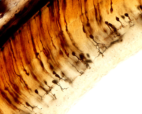
Free Images: "bestof:Origin of Vertebrates Fig 041.png Fig 41 �Retina and Optic Nerve of Petromyzon After Müller and Langerhans On the left side the Müllerian fibres and"
MÃ¥leri, genrebild - Skoklosters slott - 88958.tif
Origin_of_Vertebrates_Fig_041.png
Origin_of_Vertebrates_Fig_044.png
Origin_of_Vertebrates_Fig_047.png
Origin_of_Vertebrates_Fig_027.png
Origin_of_Vertebrates_Fig_039.png
Origin_of_Vertebrates_Fig_042.png
Origin_of_Vertebrates_Fig_032.png
Origin_of_Vertebrates_Fig_033.png
Origin_of_Vertebrates_Fig_034.png
Origin_of_Vertebrates_Fig_043.png
Origin_of_Vertebrates_Fig_031.jpg
Origin_of_Vertebrates_Fig_114.jpg
Origin_of_Vertebrates_Fig_123.jpg
Origin_of_Vertebrates_Fig_038.png
Origin_of_Vertebrates_Fig_094.png
Origin_of_Vertebrates_Fig_132.png
Origin_of_Vertebrates_Fig_154.png
Origin_of_Vertebrates_Fig_071.jpg
Origin_of_Vertebrates_Fig_116.jpg
Origin_of_Vertebrates_Fig_124.jpg
Origin_of_Vertebrates_Fig_100.png
Origin_of_Vertebrates_Fig_113.png
Origin_of_Vertebrates_Fig_120.png
Origin_of_Vertebrates_Fig_058.png
Origin_of_Vertebrates_Fig_052.png
Origin_of_Vertebrates_Fig_055.jpg
Origin_of_Vertebrates_Fig_063.jpg
Origin_of_Vertebrates_Fig_018.png
Origin_of_Vertebrates_Fig_025.png
Origin_of_Vertebrates_Fig_037.png
Origin_of_Vertebrates_Fig_054.png
Origin_of_Vertebrates_Fig_056.png
Origin_of_Vertebrates_Fig_065.png
Origin_of_Vertebrates_Fig_068.png
Origin_of_Vertebrates_Fig_069.png
Origin_of_Vertebrates_Fig_073.png
Origin_of_Vertebrates_Fig_080.png
Origin_of_Vertebrates_Fig_082.png
Origin_of_Vertebrates_Fig_083.png
Origin_of_Vertebrates_Fig_084.png
Origin_of_Vertebrates_Fig_090.png
Origin_of_Vertebrates_Fig_092.png
Origin_of_Vertebrates_Fig_093.png
Origin_of_Vertebrates_Fig_098.png
Origin_of_Vertebrates_Fig_099.png
Origin_of_Vertebrates_Fig_106.png
Origin_of_Vertebrates_Fig_119.png
Origin_of_Vertebrates_Fig_121.png
Origin_of_Vertebrates_Fig_122.png
Origin_of_Vertebrates_Fig_125.png
Origin_of_Vertebrates_Fig_126.png
Origin_of_Vertebrates_Fig_131.png
Origin_of_Vertebrates_Fig_133.png
Origin_of_Vertebrates_Fig_135.png
Origin_of_Vertebrates_Fig_140.png
Origin_of_Vertebrates_Fig_141.png
Origin_of_Vertebrates_Fig_162.png
Origin_of_Vertebrates_Fig_163.png
Origin_of_Vertebrates_Fig_057.jpg
Origin_of_Vertebrates_Fig_060.jpg
Origin_of_Vertebrates_Fig_064.jpg
Origin_of_Vertebrates_Fig_074.jpg
Origin_of_Vertebrates_Fig_115.jpg
Origin_of_Vertebrates_Fig_117.jpg
Origin_of_Vertebrates_Fig_118.jpg
Origin_of_Vertebrates_Fig_134.jpg
Origin_of_Vertebrates_Fig_019.png
Origin_of_Vertebrates_Fig_020.png
Origin_of_Vertebrates_Fig_022.png
Origin_of_Vertebrates_Fig_030.png
Origin_of_Vertebrates_Fig_053.png
Origin_of_Vertebrates_Fig_081.png
Origin_of_Vertebrates_Fig_085.png
Origin_of_Vertebrates_Fig_062.png
Origin_of_Vertebrates_Fig_078.png
Origin_of_Vertebrates_Fig_148.png
Origin_of_Vertebrates_Fig_153.png
Origin_of_Vertebrates_Fig_157.png
Origin_of_Vertebrates_Fig_158.png
Origin_of_Vertebrates_Fig_051.png
Origin_of_Vertebrates_Fig_145.png
Origin_of_Vertebrates_Fig_151.png
Origin_of_Vertebrates_Fig_011.jpg
Origin_of_Vertebrates_Fig_001.png
Origin_of_Vertebrates_Fig_103.png
Cavefaunaofnorth00pack_0183.jpg
Cavefaunaofnorth00pack_0179.jpg
Origin_of_Vertebrates_Fig_012.jpg
Origin_of_Vertebrates_Fig_013.jpg
Origin_of_Vertebrates_Fig_014.jpg
Origin_of_Vertebrates_Fig_017.jpg
Origin_of_Vertebrates_Fig_059.jpg
Origin_of_Vertebrates_Fig_101.jpg
Origin_of_Vertebrates_Fig_109.jpg
Origin_of_Vertebrates_Fig_128.jpg
Origin_of_Vertebrates_Fig_129.jpg
Origin_of_Vertebrates_Fig_136.jpg
Origin_of_Vertebrates_Fig_002.png
Origin_of_Vertebrates_Fig_003.png
Terms of Use   Search of the Day