Click Here for More Images from iStock- 15% off with coupon 15FREEIMAGES 
Cryptosporidiosis, a diarrheal disease caused by Cryptosporidium parvum protozoan. 3D illustration showing release of parasite sporozoites from oocyst inside small intestine
Human Large Intestine,Appendix in human body
Epidermis. Electron microscope micrograph showing a keratinocyte of spinous layer. The epithelial cell has a polygonal shape, central nucleus with nucleolus, cytoplasm full of keratin filament bundles, and numerous dark desmosomes crossing the intercellular spaces.
Neuroendocrine carcinoma well differentiated (carcinoid) rectum with immunostain synaptophysin
The detrusor muscle remains relaxed to allow the bladder to store urine, and contracts during urination to release urine 3d illustration
Photomicrograph of midge fly, Chironomidae family, eggs hatching. Eggs are encased in a gelatinous material that attracts debris. Live specimen. Larva about 0.5 mm long. Wet mount, 10X objective, transmitted brightfield illumination.
amazing inhabitants of the microworld under a microscope
plant Zea Stem C.S. under light mircoscope with white background
Leiden leaf c.s. under microscope
3D representation of the human body
Gastric mucosa stained with the Masson trichrome stain showing: surface epithelium, gastric pits with foveolar cells, fundic glands and muscularis mucosae.
Cardiac amyloidosis is a condition where faulty proteins build up in your heart. You can inherit this condition, or it can develop on its own (usually later in life). As faulty proteins accumulate, your heart struggles to pump, ultimately leading to heart failure and death.
Labeled Eukaryotic cell, nucleus and organelles and plasma membrane - 3d illustration
Photomicrograph of flatworm,
Bronchopulmonary segmental anatomy describes the division of the lungs into segments based on the tertiary or segmental bronchi 3d illustration
Tetis of Fish T.S. under light microscope with white background
Egg of parasitic roundworm Trichuris trichiura, or whipworm, the causative agent of trichuriasis, disease of a human large intestine, 3D illustration
Micro world
Paps smear analysis: Superficial squamous cell, metaplastic squamous cell, koilocytes cell. HPV related change
anaplastic thyroid carcinoma with heterologous differentiation (leiomyosarcoma)
Backgrounds of Characteristics Tissue Tongue, Small intestine of Cat and Tissue of Esophagus rabbit under microscope.
Photomicrograph of the ciliate Blepharisma americanum in various stages of development. Typical faint pink color. Each individual is only one cell. Live specimen. Wet mount, 10X objective, transmitted brightfield illumination.
The esophagus is the hollow, muscular tube that passes food and liquid from your throat to your stomach 3d illustration
Squash preparation of stained onion root tip cells showing various stages of mitosis. Differential interference contrast optics on a Leica microscope, Image taken with the X40 objective lens.  Depth of focus is extremely shallow (just a few microns).  Differential interference contrast results in an optical slice horizontally through the specimen providing maximum possible resolution at this magnification and good contrast. Chromatic abberation is inevitable in light microscopy, but has been kept to a minimum in this image.
Spirogyra algae (filamentous green algae) under the microscope - optical microscope x400 magnification
Micro world
3D Illustration Concept of Human Respiratory System Lungs with Diaphragm Anatomy
Fatty liver disease (steatosis) is a common condition caused by having too much fat build up in your liver. A healthy liver contains a small amount of fat. It becomes a problem when fat reaches 5% to 10% of your liver's weight.
Frogs are a diverse and largely carnivorous group of short-bodied, tailless amphibians composing the order Anura
Photomicrograph of filamentous blue-green algae, Oscillatoria species. Each band along the stand is one cell. This filament is very short probably having recently parted from a longer one. Filaments are typically many times longer. This algae is mobile, it moves and twists through the water. San Francisco Bay, California, USA. Live specimen. Wet mount, 40X objective, transmitted brightfield illumination. Note - motion blur of live specimen, very shallow depth of field, chromatic aberration and uneven focus are inherent in light microscopy.
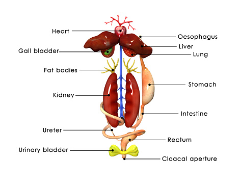
Free Images: "bestof:Origin of Vertebrates Fig 001.png Fig 1 �Arrangement of Organs in the Vertebrate A and Arthropod B Al gut; H heart; C N S central nervous system; V ventral"
151024-M-CJ278-001.jpg
Chondrostoma_nasus_(Linnaeus_1758)_Kopf_Fig_158_(Matschie_et_al._1909).svg
Complete neuron cell diagram without text.svg
1868sievers_tav3fig210.svg
1868sievers_tav3fig210_1916wolfenden_fig5.svg
@marcelosilksantos_-_vector-estampa-camiseta-vetor-em-corel-draw-vetorizao-imagens-vetorizadas-silk-screen-serigrafia-branco-e-preto-FORMANDOS-_TERCEIRÃO-_COREL-_(120).jpg
Origin_of_Vertebrates_Fig_001.png
Origin_of_Vertebrates_Fig_167.png
Origin_of_Vertebrates_Fig_072.png
Origin_of_Vertebrates_Fig_003.png
Origin_of_Vertebrates_Fig_112.png
Origin_of_Vertebrates_Fig_168.png
Origin_of_Vertebrates_Fig_160.png
Origin_of_Vertebrates_Fig_156.png
Origin_of_Vertebrates_Fig_159.png
Origin_of_Vertebrates_Fig_021.png
Origin_of_Vertebrates_Fig_071.jpg
Origin_of_Vertebrates_Fig_124.jpg
Origin_of_Vertebrates_Fig_040.png
Origin_of_Vertebrates_Fig_058.png
Archi-mollusc_by_Herdman.jpg
Origin_of_Vertebrates_Fig_019.png
Origin_of_Vertebrates_Fig_044.png
Origin_of_Vertebrates_Fig_125.png
Origin_of_Vertebrates_Fig_166.png
Origin_of_Vertebrates_Fig_074.jpg
Origin_of_Vertebrates_Fig_045.png
Origin_of_Vertebrates_Fig_094.png
Origin_of_Vertebrates_Fig_054.png
Origin_of_Vertebrates_Fig_084.png
Origin_of_Vertebrates_Fig_103.png
Origin_of_Vertebrates_Fig_119.png
Origin_of_Vertebrates_Fig_144.png
Origin_of_Vertebrates_Fig_146.png
Origin_of_Vertebrates_Fig_147.png
Origin_of_Vertebrates_Fig_148.png
Origin_of_Vertebrates_Fig_150.png
Origin_of_Vertebrates_Fig_152.png
Origin_of_Vertebrates_Fig_063.jpg
Origin_of_Vertebrates_Fig_118.jpg
Origin_of_Vertebrates_Fig_134.jpg
Origin_of_Vertebrates_Fig_020.png
Origin_of_Vertebrates_Fig_034.png
Origin_of_Vertebrates_Fig_068.png
Origin_of_Vertebrates_Fig_080.png
Origin_of_Vertebrates_Fig_091.png
Origin_of_Vertebrates_Fig_110a.png
Origin_of_Vertebrates_Fig_120.png
Origin_of_Vertebrates_Fig_139.png
Origin_of_Vertebrates_Fig_060.jpg
Origin_of_Vertebrates_Fig_109.jpg
Origin_of_Vertebrates_Fig_026.png
Origin_of_Vertebrates_Fig_046.png
Origin_of_Vertebrates_Fig_047.png
Origin_of_Vertebrates_Fig_062.png
Origin_of_Vertebrates_Fig_078.png
Origin_of_Vertebrates_Fig_082.png
Origin_of_Vertebrates_Fig_083.png
Origin_of_Vertebrates_Fig_090.png
Origin_of_Vertebrates_Fig_110c.png
Origin_of_Vertebrates_Fig_157.png
Origin_of_Vertebrates_Fig_158.png
Principles_of_Psychology_(James)_v1_p21.png
Life_in_Motion_Fig_80.png
The_Quarterly_journal_of_the_Geological_Society_of_London_(13205249514).jpg
BritishTunicataPlate3.jpg
Surface projections of the organs of the trunk.png
Image_from_page_46_of_"Relics_of_primeval_life,_beginning_of_life_in_the_dawn_of_geological_time"_(1897).jpg
Origin_of_Vertebrates_Fig_011.jpg
Origin_of_Vertebrates_Fig_012.jpg
Origin_of_Vertebrates_Fig_013.jpg
Origin_of_Vertebrates_Fig_014.jpg
Origin_of_Vertebrates_Fig_017.jpg
Origin_of_Vertebrates_Fig_031.jpg
Origin_of_Vertebrates_Fig_055.jpg
Origin_of_Vertebrates_Fig_057.jpg
Origin_of_Vertebrates_Fig_059.jpg
Origin_of_Vertebrates_Fig_064.jpg
Origin_of_Vertebrates_Fig_101.jpg
Origin_of_Vertebrates_Fig_114.jpg
Origin_of_Vertebrates_Fig_115.jpg
Origin_of_Vertebrates_Fig_116.jpg
Origin_of_Vertebrates_Fig_117.jpg
Origin_of_Vertebrates_Fig_123.jpg
Origin_of_Vertebrates_Fig_128.jpg
Origin_of_Vertebrates_Fig_129.jpg
Origin_of_Vertebrates_Fig_136.jpg
Origin_of_Vertebrates_Fig_002.png
Origin_of_Vertebrates_Fig_004.png
Origin_of_Vertebrates_Fig_005.png
Origin_of_Vertebrates_Fig_006.png
Origin_of_Vertebrates_Fig_007.png
Origin_of_Vertebrates_Fig_008.png
Origin_of_Vertebrates_Fig_009.png
Origin_of_Vertebrates_Fig_010.png
Origin_of_Vertebrates_Fig_015.png
Origin_of_Vertebrates_Fig_016.png
Origin_of_Vertebrates_Fig_018.png
Origin_of_Vertebrates_Fig_022.png
Origin_of_Vertebrates_Fig_023.png
Terms of Use   Search of the Day