Click Here for More Images from iStock- 15% off with coupon 15FREEIMAGES 
Dna structure on science background. 3d illustration
Molecular model of fullerene (c60)
The Raf/MEK/ERK (MAPK) propagates growth signals in the cell. Misregulation can lead to cancer.\nThe red dots on the active EGFR are phosphorylated tyrosines. PDB entries: 1egf, 1nql, 1m17, 2jwa, 3njp, 2gs6, 1gri, 1xd2, 3ksy, 5p21, 6xi7, 6q0j, 2y4i,
What micro plastic is made of - Chemical ingredients.\nClose up of microplastic on finger concept of water pollution and global warming
Ovarian follicles. Light microscopy, hematoxylin and eosin stain, magnification 200x and 3D illustration
background
Nucleosome structure. Structure of an oligonucleosome, showing the packaging of DNA in chromosomes. 3D illustration.
Fetus with dna 3D rendering
High resolution 3d render of cells
The three-dimensional structure of proteins is represented in a variety of conformations. This image is based on the ribbon structure of a protein. It is suitable for biology notebooks and book covers.\nThis image has not been created by AI.
B-chain pentamer and A-chain subunit (green), 3D cartoon and Gaussian surface model, white background
Molecular structure formula. Concept. 3D Render
DNA close-up, DNA Molecule Model, DNA Strands, DNA animation
3d rendering of Easter eggs background
Molecular genesis of Huntington's disease, 3D illustration. Expansion of the CAG trinucleotide sequence in the htt gene causes production of mutated Huntingtin protein leading to neurodegeneration
anaplastic thyroid carcinoma with heterologous differentiation (leiomyosarcoma)
Group of blue plant or algae organism cells on dark background, micro view. Medical and microbiological scientific research illustration. 3D rendering
plant Leaf of Nerium indicum T.S. under light mircoscope with white background
Aerial view of loyster farm by Ardara, County Donegal - Ireland.
Conceptual global crisis of Covid-19 virus epidemic. Covid-19 virus illustration downloaded from CDC than layered and manipulated. for more information please visit Centers for Disease Control and Prevention web site: https://www.cdc.gov/media/subtopic/images.htm
The glucagon like peptide-1 receptor (GLPr) is a G proteincoupled receptor (GPCR) that mediates the action of GLP-1, a peptide hormone. The activated receptor  has a strong effect  on the management of type 2 diabetes mellitus and obesity, including glucose homeostasis and regulation of gastric motility and food intake.
Photomicrograph of filamentous blue-green algae, Oscillatoria species. Each band along the stand is one cell. This filament is very short probably having recently parted from a longer one. Filaments are typically many times longer. This algae is mobile, it moves and twists through the water. San Francisco Bay, California, USA. Live specimen. Wet mount, 40X objective, transmitted brightfield illumination. Note - motion blur of live specimen, very shallow depth of field, chromatic aberration and uneven focus are inherent in light microscopy.
Fungal infection on a human hand. Ringworm, tinea manuum and close-up view of dermatophyte fungi Trichophyton mentagrophytes, 3D illustration
Human embryonic stem cells
Bronchitis and alveolitis in lung of horse infected with equine influenza virus, 200x magnification.
3d rendering of transparent molecule shape on green color background.
Nodularia sp. toxic algae under microscopic view, nitrogen-fixing cyanobacteria, cyanotoxin, harmful to humans, x40
Myxoid liposarcoma.  Myxoid/round cell liposarcoma, or MRCLS, is one of several types of liposarcoma. Liposarcoma is a rare cancer that grows in the cells that store fat in the body. MRCLS usually grows in the arms and legs. These tumors grow slowly, and they can spread to other parts of the body
From left to right: the NMDA and  AMPA receptors transport calcium cations into neurons after being activated by the neurotransmitter glutamate and  the GABA receptor (right) transport chloride anions after the activation by gamma aminobutyrate. PDB entries: 6wht,  3kg2, 6d6u
Tetis of Fish T.S. under light microscope with white background
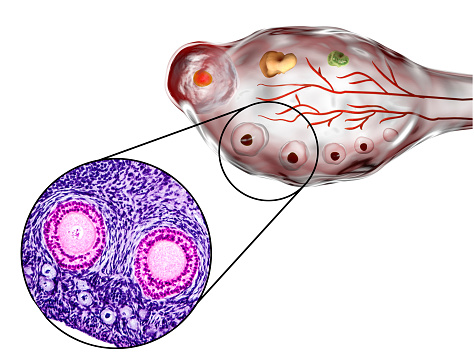
Free Images: "bestof:Oogenesis-rus.png en Diagram showing the reduction in number of the chromosomes in the process of maturation of the ovum Figure 5 from Gray's Anatomy ru"
Gray5.svg
Gray5_de.svg
Gray5_nl.svg
Gray5_-_es.svg
Gray5-ja.svg
Gray's_7_(ovum_maturation).svg
Diagram of swine influenza symptoms EN.svg
Diagram of influenza infection symptoms EN.svg
PD Diagram of swine flu symptoms EN.svg
Circulatory System ru.svg
Gray1046_pl.svg
Gray1193_pl.svg
Brain diagram fr.svg
Brain_diagram_ja.svg
Brain_diagram_without_text.svg
Gray769-pt.svg
Circulatory System en edited.svg
Circulatory System en.svg
Rail Tracks map Kintetsu Uehonmachi Station VII en.svg
Rail Tracks map Kintetsu Uehonmachi Station III.svg
Rail Tracks map Kintetsu Uehonmachi Station IV.svg
Rail Tracks map Kintetsu Uehonmachi Station VII.svg
George_Stubbs_-_A_Comparative_Anatomical_Exposition_of_the_Structure_of_the_Human_Body_with_that_of_a_Tiger_and_a_Co..._-_Google_Art_Project_(2349640).jpg
Crossover genes zh.svg
WP Offline Proccess es.svg
WP Offline Proccess en.svg
Trilobite sections-ru.svg
Interpreted Code Flow Diagram.svg
Compiled Code Flow Diagram.svg
Blood Compatibility.svg
Trilobite sections numbered.svg
Trilobite cranidium numbered.svg
Trilobite cranidium-en.svg
CutFill.svg
Tree secondary components diagram.svg
Culex_pipiens_diagram_en.svg
Outer hair cell and Deiter's cell.svg
Wignspan Diagram.svg
Trilobite cephalon areas numbered.svg
Trilobite cephalon areas-en.svg
Ant worker morphology (ja).svg
Culex pipiens diagram es.svg
Culex pipiens diagram uk.svg
HEPA Filter diagram en.svg
TRNA ribosomes diagram ru.svg
Culex pipiens diagram fr.svg
Culex pipiens diagram zh.svg
Brain_diagram_pl.svg
Gray399_es.svg
P-v diagrams elementary evolutions liquids-vapors.svg
Gray834 pl.svg
Human skull side details right pl.svg
Human skull side suturas pl.svg
Human skull side details pl.svg
Gray399.svg
Gray1193.svg
Gray1167_pl.svg
Стомах.svg
Cross section jellyfish en.svg
Gray728.svg
Gray1216_modern_locations.svg
Complete diagram of a human spermatozoa en.svg
Jellyfish cross section numbered.svg
TRNA ribosomes diagram pl.svg
Gray834.svg
Gray770-pt.svg
US timeline. Heroin deaths.svg
Gray1046.svg
Lobes_of_the_brain_el.svg
Temporo-parietal_junction_ja.svg
US timeline. Prescription opioid pain reliever deaths.svg
Voting-age.svg
Cross section jellyfish ru.svg
Gray769-en.svg
Venous system en.svg
Arterial System en.svg
Circulatory System fr.svg
Arterial System fr.svg
Gray834_es.svg
Gray799.svg
Gray1236_pl.svg
Gray769-es.svg
Complete neuron cell diagram numbered large numbers.svg
Circulatory System no tags.svg
Rail Track diagram of Tambabashi Station 1967 03 en.svg
Gray1235_pl.svg
Gray814_pl.svg
Complete diagram of a human spermatozoa fr.svg
Gray67.svg
Human arm bones diagram.svg
Complete neuron cell diagram ru.svg
Scheme ant worker anatomy-en.svg
Sylvester-square.svg
Complete diagram of a human spermatozoa pt.svg
Joseph_Walter_-_Shipping_off_Bristol.jpg
Complete diagram of a human spermatozoa pl.svg
Rail Tracks map Kintetsu Uehonmachi Station VI en.svg
Rail Track diagram of Tambabashi Station 1967 03 ja.svg
Judith_with_the_Head_of_Holofernes_by_Cristofano_Allori_(detail).jpg
George_Stubbs_-_A_Comparative_Anatomical_Exposition_of_the_Structure_of_the_Human_Body_with_that_of_a_Tiger_and_a_Co..._-_Google_Art_Project_(2348692).jpg
Terms of Use   Search of the Day