Click Here for More Images from iStock- 15% off with coupon 15FREEIMAGES 
Computer generated image of kidney cross section showing the kidney interior with renal arteries and veins, with anatomy labels on a white background.
plant Flower bud of Lilium brownii var.viridulum T.S.\nunder light mircoscope with white background
A 3D medical illustration of the sacrum bone isolated on a white background, showcasing its shape, structure, and anatomical features. Back view
Human Internal System .Anatomical model  mannequin
Female anatomy. Doctor showing anatomical model of female reproductive system
The image depicts a near median section through the nose and nasal passages. Details include nasal cavity, soft and hard palate, uvula, eustachian tube and pharyngeal tonsil. White background.
Doctor with human Colon anatomy model. Colonic disease, Large Intestine, Colorectal cancer, Ulcerative colitis, Diverticulitis, Irritable bowel syndrome, Digestive system and Health concept
skin science 3d
Excretion is the process by which waste products of metabolism and other non-useful materials are eliminated from an organism.
Gynecologist holding and showing an ovary model
Leiden leaf c.s. under microscope
plant rape flower cross section under light mircoscope with white background
Digitally restored from a late 19th Century encyclopedia.
Golden Yen Symbol
Internal structure of an animal cell, 3d rendering. Section view. Computer digital drawing.
3D representation of the human body
Celiac or coeliac intestine disease anatomy medical concept with normal villi and damaged small bowel lining as an autoimmune disorder of the digestion system with colon and stomach as a 3D illustration.
Doctor holding and showing  human Inner ear model of auditory system
The fifth lumbar spine vertebrae (L5) is part of the greater lumbar region 3d illustration
Human anatomy model isolated on white background. Photographs in sequence. Photo n. 2.
3D Illustration Concept of Human Circulatory System Heart Anatomy
axial lung window chest ct scan 50 years old patient with chest pain showing  bowel  dan gaster visible in  left chest and displacement of the mediastinum to the right
Computational tomography image of the abdominal and pelvic regions.
Enlarged Prostate organ gland as Benign Prostatic Hyperplasia as a urology medical illustration concept for part of the male reproductive anatomy and lower urinary tract symptoms.
Doctor holding a  human head model
A flower, sometimes known as a bloom or blossom, is the reproductive structure found in flowering plants
Human small and large intestines internal organ 3D design element.
Isolated kidney cross section showing the kidney interior with renal arteries and veins.
MRI scanning of the human body skeletal, muscular and nervous systems with foot and knee region view.
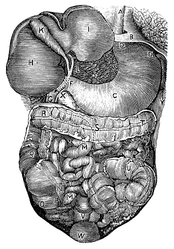
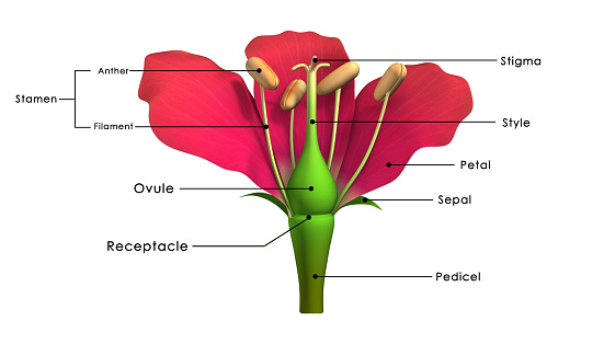
Free Images: "bestof:Oligochaeta anatomy 2.svg Diagrama Anatómico de un Oligoqueto 1 - lumen intestinal 2 - tiflosolio 3 - quetas 4 - cutícula 5 - vasos sanguíneos 6 -"
Terms of Use   Search of the Day