Click Here for More Images from iStock- 15% off with coupon 15FREEIMAGES 
Components of Eukaryotic cell, nucleus and organelles and plasma membrane - 3d illustration
Cell Membrane Phospholipid Structure,Phospholipid bilayer of the cell membrane,Lipid bilayer,lipid bi-layer cell anatomy
Organelles inside an Eukaryote or eukaryotic cell. 3d illustration
3d cell organelle - nucleus (Chromatin,  Rough Endoplasmic Reticulum, Nucleus Shell, Nucleoplasm, Nucleolus)
Detailed 3D illustration of hypothalamic nuclei, showcasing the brain's vital control center for various physiological functions.
Mid-sagittal section showing the anatomy of the pelvis and testis. The model shot in studio environment. Isolated on white background.
Adipose tissue human, Smooth muscles human, Bone human and Striated (skeletal) muscle human under the microscope in Lab.
3D illustration showing layers of the gastrointestinal tract isolated on white
Human eye anatomy on scientific background. 3d illustration
plant Pine Stem C.S. under light mircoscope with white background
Internal structure of an animal cell, 3d rendering. Section view. Computer digital drawing.
Mesenchymal dysplasia. Site: Placenta
3d rendering of biological animal cell with organelles cross section isolated on white. Animal cell with placed text annotations to all organelles
Human embryonic stem cells
Organic cell. Virus, germ or bacteria. Isolated on white background. 3d render
Close-up shooting of a green agate stone mineral surface isolated on white background. Its a macro shot shows nature beautiful details with back lit. Agate is a kind of natural gemstone. Abstract pattern. Good for Wallpaper texture background. Purple
Labeled Eukaryotic cell, nucleus and organelles and plasma membrane - 3d illustration
plant rape flower cross section under light mircoscope with white background
The kidneys are two bean-shaped organs that filter your blood3d illustration Your kidneys are part of your urinary system 3d illustration
The female reproductive system consists of internal and external organs. It creates hormones and is responsible for fertility, menstruation and sexual activity.
A microscope slide showing a stained section of a human bowel, or small intestine.  It was taken through microscope lens that has a central focus through a thin layer of glass.
Showing Light micrograph Type of Tissue Human under the microscope in Lab.
3d animal cell on black background
brown fat cell also called brown adipose tissue side to side with white fat cell 3d rendering
Rosai–Dorfman disease (RDD) is a rare proliferative histiocytic disorder of unknown etiology. RDD typically presents with generalized lymphadenopathy and polymorphic histiocytic infiltration of the lymph node sinuses; however, occurrences of extranodal soft tissue RDD may rarely occur when masquerading as a soft tissue sarcoma.
Internal structure of an animal cell, 3d rendering. Section view. Computer digital drawing.
Oviduct T.S. under light microscope with white background
Human eye anatomy.inner structure.3d illustration
Abstract purple stone background, nature pattern.
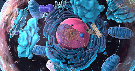
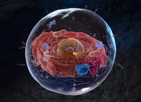
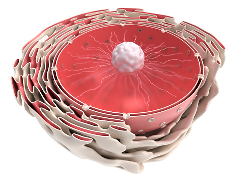
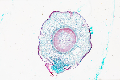
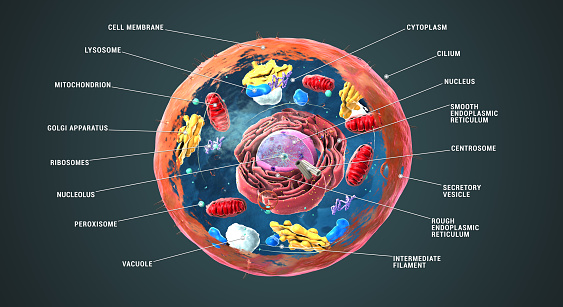
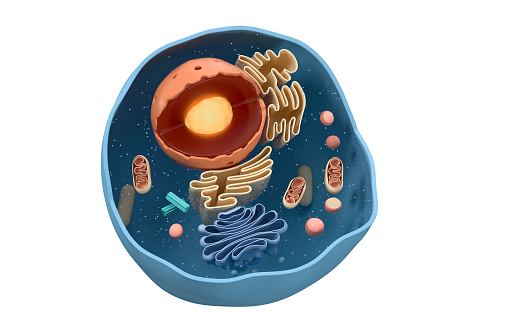
Free Images: "bestof:Nucleus ER golgi ex.jpg Secretory pathway diagram including nucleus endoplasmic reticulum and Golgi apparatus Nuclear membrane Nuclear pore Rough endoplasmic"
Plant cell structure svg labels.svg
Animal Cell.svg
Endomembrane system diagram en.svg
Plant cell structure-en.svg
Plant cell structure svg 2.svg
Thyroid hormone synthesis-it.svg
Thyroid hormone synthesis.svg
Diagram human cell nucleus numbered version.svg
Nucleus ER.svg
Animal Cell Unannotated.svg
Diagram human cell nucleus.svg
Diagram human cell nucleus cs.svg
Nuclear fission.svg
Apicomplexa structure.svg
ProteinQS.svg
Golgi apparatus (borderless version)-pt.svg
atom-nucleus-nuclear-atom-nucleus-153152.svg
nuclear-power-atom-atomic-nucleus-598820.jpg
atomic-nucleus-nuclear-energy-467254.svg
atom-atomic-nucleus-nuclear-power-1015454.jpg
atom-science-nuclear-nucleus-152980.svg
Nuclear predetonation.svg
atom-nucleus-atomic-nucleus-atom-296747.svg
orbit-orbital-atom-atomic-nucleus-467260.svg
atom-electron-neutron-nuclear-power-1222514.jpg
atom-electron-neutron-nuclear-power-1222516.jpg
atom-electron-neutron-nuclear-power-1222512.jpg
atom-electron-neutron-nuclear-power-1222513.jpg
atom-electron-neutron-nuclear-power-1222511.jpg
atom-physics-atomic-nucleus-neutron-618331.jpg
atom-nuclear-power-symbol-proton-1015667.jpg
atom-nuclear-power-symbol-proton-1015673.jpg
atom-nuclear-power-symbol-proton-1015707.jpg
Lipid raft organisation scheme.svg
Euglenid pellicula scheme.svg
ThalamicNuclei.svg
atom-nuclear-chemistry-education-42601.svg
Nuclear fusion.svg
hands-protect-protection-father-1651146.jpg
hands-protect-protection-father-1651147.jpg
silhouette-nuclear-power-plant-1312223.png
silhouette-nuclear-power-plant-1311985.png
hands-protect-protection-father-598145.jpg
hands-protect-protection-father-598146.jpg
Hydrogen bomb.svg
Atomic bomb.svg
Little Boy internal diagram.svg
Little Boy internal diagram (no labels).svg
Binding energy curve - common isotopes DE.svg
Binding energy curve - common isotopes-CZ.svg
Binding energy curve - common isotopes es.svg
Binding energy curve - common isotopes fi.svg
Binding energy curve - common isotopes.svg
Binding energy curve - all.svg
BWR Mark I Containment sketch with downcomers.svg
Nucleus ER golgi ex.jpg
Nucleus ER golgi.jpg
Nucleus ER.png
Thyroid hormone synthesis.png
Pearl_Bodies04.jpg
Diagram human cell nucleus serbian nuclear pore.PNG
Mammalian pancreatic tissue, nuclear pores and rough endoplasmic reticulum.jpg
Pancreatic acinar cells - TEM.jpg
Clara cell lung - TEM.jpg
Diagram human cell nucleus serbian nuclear envelope.PNG
Cell plate miguelferig.png
HLA-DQ protein.PNG
Linking-Human-Diseases-to-Animal-Models-Using-Ontology-Based-Phenotype-Annotation-pbio.1000247.g006.jpg
Nfowleri_lifecycle.gif
O.Hertwig1906Fig2-6.jpg
Spindle_with_Four_Poles_(8744518858).jpg
BWR Mark I Containment sketch with downcomers.png
BWR Mark I Containment sketch primary.png
Diagram of nuclear uptake of protein.jpg
PBB Protein ERP29 image.jpg
Nuclear dens.png
Nuclear Torn bandpic.jpg
Isotopes and half-life.PNG
Macrophage in the alveolus Lung - TEM.jpg
A red blood cell in a capillary, pancreatic tissue - TEM.jpg
ThalamicNuclei.png
Endomembrane system ar.png
Nuclear components ru.jpg
Diseases of Swine 23-3.png
Type ii nuclear receptor action.png
Prolatelow2.png
Prolatehigh.png
Nuclear receptor action.png
Radiolaria_(Challenger)_Plate_065.jpg
Isotopes and half-life 1.PNG
Origin_of_Vertebrates_Fig_042.png
Radiolaria_(Challenger)_Plate_002.jpg
BWR_Mark_I_Containment,_diagram.png
Annihilation.png
BWR Mark I Containment sketch.png
Littleboy inner1 info.png
Terms of Use   Search of the Day