Click Here for More Images from iStock- 15% off with coupon 15FREEIMAGES 
A carboxypeptidase is a protease enzyme that hydrolyzes a peptide bond at the carboxy-terminal end of a protein or peptide. 3D cartoon model, secondary structure color scheme, PDB 2v77, white background.
The three-dimensional structure of proteins is represented in a variety of conformations. This image is based on the ribbon structure of a protein. It is suitable for biology notebooks and book covers.\nThis image has not been created by AI.
Bacterial multidrug efflux pumps (E-CmeB) expel antibiotics  from inside the bacterium (bottom): ciprofloxacin (yellow), chloramphenicol (violet),  ampicillin (red).
A ball and stick model of Human Insulin. Isolated on black.
3d rendered Gastric inhibitory polypeptide (GIP, glucose-dependent insulinotropic peptide) endocrine protein hormone, 3D illustration
Inside protein molecule
Proteasomes are molecular machines for breaking down proteins called proteolysis, only target proteins that have been marked for destruction called ubiquitin to the target protein, 3d rendering
Top view of various kinds of vegan protein sources like tofu, tempeh, soy beans, soy milk, mushrooms, wakame, lentils, peanuts, spinach and chick peas. All the objects are on a gray bluish backdrop. Studio shot taken with Canon EOS 6D Mark II and Canon EF 24-105 mm f/4L
3D cartoon and Gaussian surface models, PDB 2gjx, white background
Covid-19 Coronavirus Variants Omicron Delta Gamma Beta Alpha Pandemic Virus Disease Mutation
The crystal structure of the tumor marker protein. The 3D model of the biological macromolecule.
Abstract large group of metallic sphere particles background. Metaverse background concept.
A ribbons model of Human Growth Hormone, also known as Somatotropin, with a translucent surface model.
orange: RNA primer; red: RNA template; light blue: proofreading riboexonuclease; mid blue: helicase; dark blue: RNA directed RNA polymerase; violet: non structural proteins (NSPs).
Variety of healthy vegan, plant based protein source and body building food. Tofu soy beans tempeh, green vegetables, nuts, seeds, quinoa oat meal and spirulina. View from above
Cell Membrane Phospholipid Structure,Phospholipid bilayer of the cell membrane,Lipid bilayer
3D cartoon and Gaussian surface models, chain id color scheme, PDB 3omv, white background
Overhead view of a large group of food with high content of healthy proteins arranged side by side on black background making a banner. The composition includes salmon beef fillet, chicken breast, eggs, yogurt, mussels, chick peas, pistachios, cheese, brown lentild, beans, shrimps, canned tuna, pumpkin seeds, soybeans among others. High resolution 42Mp studio digital capture taken with SONY A7rII and Zeiss Batis 40mm F2.0 CF lens
Foods containing natural magnesium (Mg). Healthy food concept. Top view, copy space
DNA A-T Pair
c-Myc and Max transcription factors bound to DNA. 3D illustration. Atoms shown as spheres with conventional color coding.
A ball and stick + ribbons model of beta-secretase - a protein that is important in forming healthy peripheral nerve cells, but is also involved in the onset of Alzheimer's disease.
Collagen protein molecule. Molecular model. 3D rendering. Illustration
Shiny artificial molecular structure in many colors. Horizontal composition, isolated on black background.
3D cartoon and Gaussian surface models, PDB 6vg8, white background
Phospholipid bilayer membrane structures that make up living cells and their contents. This is an image of membrane proteins in the membrane.\nThis image, which looks like a plastic model, evokes biology and biochemistry. It can be used as a cover for magazines, textbooks and presentation materials.\nThis image was not created by AI.
p53 (blue) tagged with ubiquitin (yellow) is degraded into small peptides by a proteasome (violett). p53 prevents cancer formation and acts as a guardian of the genome. Mutations in the p53 gene contribute to about half of the cases of human cancer.
High angle view of a group of food with high calcium content arranged at the right leaving copy space at the left. The composition includes dairy products souch milk, yogurt, butter, various cheeses, mozzarella cheese, eggs, sliced orange, broccoli, mixed beans, sardines, almonds among others. High resolution 42Mp studio digital capture taken with SONY A7rII and Zeiss Batis 40mm F2.0 CF lens
raw salmon, meat and chicken breasts near green vegetables isolated on grey, ketogenic diet menu
Dna 3D rendering, NO AI create
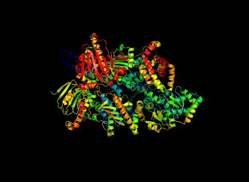
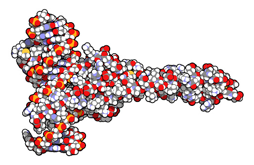
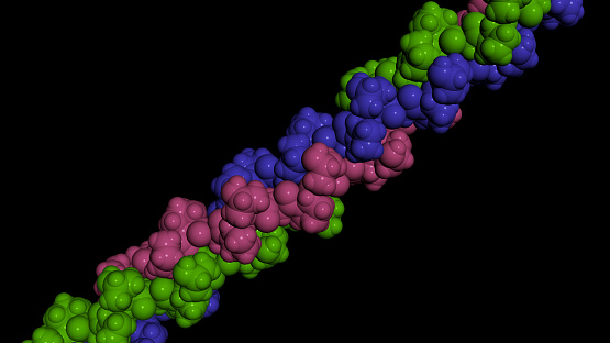
Free Images: "bestof:Main protein structure levels pl.svg protein structures Struktury budowy białek Main_protein_structure_levels_en svg Translation M Komorniczak"
Main protein structure levels pl.svg
Main protein structure levels pl - secondary structure.svg
Main protein structure levels zh.svg
Main protein structure levels bg.svg
Main protein structure levels zh (zh-tw).svg
Main protein structure levels en.svg
Main protein structure levels fr.svg
Main protein structure levels uk.svg
Main protein structure levels gl.svg
Penis_pl.svg
Aa structure function.svg
DNA replication pl.svg
2ph1 pdb gallery nucleotide binding-protein-2 (MinD homolog).svg
Complete diagram of a human spermatozoa pl.svg
Molbio-Header-2.svg
Stat domain structure (zh-cn).svg
Human arm bones diagram PL.svg
TRNA ribosomes diagram pl.svg
Molbio-Header.svg
Thyroid hormone synthesis-it.svg
Eukaryotic cilium diagram en.svg
SJA-WorldMap.svg
Europe Location Georgia.svg
Europe Location Azerbaijan.svg
Europe Location Armenia.svg
Magaya client distribution.svg
Countries adopting Metric System.svg
IPTV-Countries.svg
Main protein structure levels bg.png
PDB 1cmw EBI.jpg
PDB 1df9 EBI.jpg
PDB 1bef EBI.jpg
PDB 2qid EBI.png
Steroid receptor.png
GHCS protein structure.jpg
Mercuric reductase protein structure.jpg
Possible SYNE1 protein structure.png
G-Protein.png
Bcl-2 protein.png
Bak protein.png
Bax protein.png
Helice alpha spire 0.png
PDB 1l6l EBI.jpg
PDB 1g40 EBI.jpg
PDB 1y8e EBI.jpg
PDB 1rid EBI.jpg
PDB 1g44 EBI.jpg
PDB 2hr0 EBI.png
PDB 2a01 EBI.png
PDB 2ou1 EBI.png
4f0a bio r 500.jpg
Apolipoprotein A1 2A01.gif
IL17F 1JPY.png
1ugm bio r 500.jpg
2ykp bio r 500.jpg
ACD-TPP1 rasmol.png
MDR3 3g5u.png
Reverse Transcriptase 1HMV.png
Thymosin 1HJ0.png
HLA B^1501.png
P35 structure by Fisher 1999, PDB 1P35.jpg
Sf caspase-1 structure by Ni 2006, PDB 2NN3.jpg
Human_eIF1A.jpg
Sulfotransferase SULT1A3.png
ECOR1 Crystal Structure.rsh.png
LSm 1I5L 1.JPG
Glutamate carboxypeptidase II.png
2BTQ.jpg
1vyb bio r 500.jpg
Human_eIF1.jpg
2gpq_asym_r_500.jpg
ZNF184_tandem_19.png
Terms of Use   Search of the Day