Click Here for More Images from iStock- 15% off with coupon 15FREEIMAGES 
Top view healing chaga mushroom on old birch trunk close up. Red parasite mushroom growth on tree. Bokeh background.
Close shot of an Oudemansiella mucida, commonly known as porcelain fungus.
White Oyster Mushrooms growing on a decaying log in a forest. High quality photo
Horizontal high angle photo of beautiful blue, white and brown striped Turkey Tail Fungus growing on an old tree stump
Large mound of bright golden yellow and green variegated foliage of Spindle Euonymus 'Emerald 'n' Gold'
Photomicrograph of Volvox globator, a green algae in various stages of development. Dark green spots are daughter colonies forming inside of parent colony. Live specimen. Wet mount, 10X objective, transmitted brightfield illumination.
Trametes pubescens
Backgrounds of Characteristics and Different shaped Colony of Bacteria and Mold growing on agar plates from Soil samples for education in Microbiology laboratory.
Golden rain or laburnum tree in bloom on the outskirts of the town of Schalkenmehren in Germany
Sunlit underside of hexagonal-pored polypore (a bracket fungus or mushroom) on dead beech tree, autumn, in the Connecticut woods. Scientific name: Polyporus alveolaris.
Representatives of the Lemnaceae, the duckweed family, are well-adapted for use as bioindicators for testing soil and water for toxic substances.  Their rapid multiplication and the simplicity of their anatomy are important advantages for this use.
Candida albicans grown on chromogenic agar plates. Candida albicans, a fungus naturally present in small quantities on the body, inhabits areas such as the mouth, skin, and intestines. When the balance with healthy bacteria is disrupted, Candida can lead to infections like thrush and vaginal yeast infections. These infections are frequent and typically addressed with antifungal medications.
Early spring yellow flowering groundcover plant Stonecrop
Close-up shot of a petri dish for checking an antibiotic in the laboratory.
Yellow slime mold, also called dog vomit slime mold, Physarum polycephalum, growing over a fallen log in the Belding Wildlife Management Area of Vernon, Connecticut.
Bracket fungus
Top view soil microorganisms Nutrient agar in plate on black background.
Autumn photos in Denmark
shrub of cherry laurel, flowers and leaves, Prunus laurocerasus; Rosaceae
Candida is the most common fungal pathogen, with Candida albicans and Candida tropicalis accounting for 90% of the infections.
Turkey tail Fungus - Trametes versicolor.
Filemantous Fungi grownon CHROMagar
Chicken of the Woods or Sulphur Shelf  Mushrooms Growing on the Tree Trunk
In the row. Trichoderma and Sclerotinia in the laboratory in the special rounded containers.
False coral fungus (Tremellodendron schweinitzii) in the Connecticut woods, midsummer. Despite its appearance and leathery texture, it is a jelly fungus. It grows on the ground near hardwood trees and gets nutrients from their roots.
A petri dish with common yeast and bacteria colonies.
Forest in the countryside in late summer / early autumn
Branch of Buxus sempervirens with dry leaves in autumn
Autumn grass and herbs in the afternoon light in autumn.
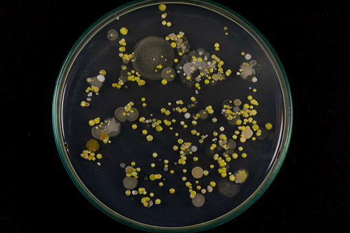
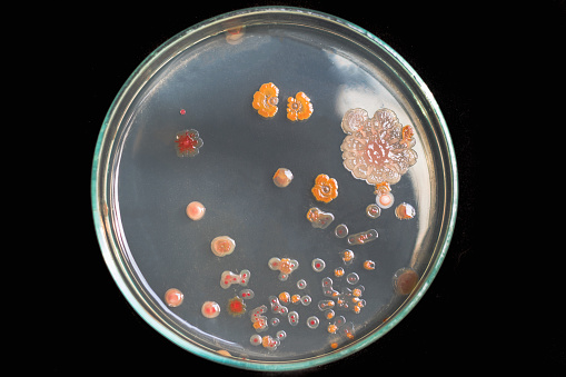
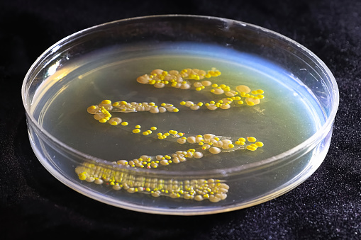
Free Images: "bestof:Madurella grisea PHIL 4158 lores.jpg This image shows a plate culture growing the fungus Madurella grisea Georgia isolate Some Madurella spp are a cause of"
Madurella_grisea_PHIL_4158_lores.jpg
Trichophyton_mariatii_PHIL_3053_lores.jpg
Acremonium_falciforme_PHIL_4167_lores.jpg
Trichophyton_terrestre_PHIL_4300_lores.jpg
Trichophyton_rubrum_var._rodhaini_PHIL_4248_lores.jpg
Plate_culture_of_Exserohilum_rostratum_PHIL_3786_lores.jpg
Trichophyton_mentagrophytes_PHIL_2929_lores.jpg
Conidia_and_conidiophores_of_the_fungus_Acremonium_falciforme_PHIL_4168_lores.jpg
Chain_of_conidia_of_a_Alternaria_sp._fungus_PHIL_3963_lores.jpg
Chain_of_conidia_of_a_Alternaria_sp._fungus_PHIL_3962_lores.jpg
Conidia-laden_conidiophores_of_Phialophora_verrucosa_PHIL_4238_lores.jpg
Microconidia_of_the_fungus_Trichophyton_mariatii_PHIL_3050_lores.jpg
Conidiophore_of_the_fungus_Bipolaris_hawaiiensis_PHIL_4338_lores.jpg
Tuberculate_macroconida_of_the_Jamaican_isolate_of_Histoplasma_capsulatum_PHIL_4023_lores.jpg
Terminal_chlamydospores_of_the_fungus_Microsporum_audouinii_PHIL_3054_lores.jpg
Macroconidia_of_the_dermatophytic_fungus_Epidermophyton_floccosum_PHIL_4207_lores.jpg
The_fine_branching_tubes_of_the_fungus_Exserohilum_rostratum_PHIL_3784_lores.jpg
Rice_blast.jpg
Conidiophores_and_conidia_of_the_fungus_Sporothrix_schenckii_PHIL_4208_lores.jpg
Tinea_versicolor_due_to_Malassezia_furfur_PHIL_2914_lores.jpg
Cataracts_due_to_Congenital_Rubella_Syndrome_(CRS)_PHIL_4284_lores.jpg
Phlebotomus_sp._6274_lores.jpg
Plasmodium_falciparum_nephrosis_edema_PHIL_3894_lores.jpg
Pneumocystis_carinii_PHIL_2998_lores.jpg
Ringworm_due_to_Trichophyton_rubrum_PHIL_2909_lores.jpg
Haemophilus_ducreyi_5811_lores.jpg
Plasmodium_falciparum_rings_form_parasites4885_lores.jpg
Plasmodium kierto.png
Cryptococcosis_of_lung_in_patient_with_AIDS_Methenamine_silver_stain_963_lores.jpg
MRSA_SEM_7821_lores.jpg
FMIB_40044_Potamogeton_heterophyllus_(16-17);_Potamogeton_perfoliatus_(18-19).jpeg
Dam_i_högreståndsdräkt,_1400-tal_-_Nordiska_Museet_-_NMA.0041172.jpg
FMIB_47814_Marennes_(La_Tremblade)_View_of_the_claires_for_greening_the_oysters.jpeg
FMIB_42225_General_view_of_the_plant_from_the_outer_rear_corner_In_foreground_one_of_the_cars_shows_the_propeller_shaft_and_faint.jpeg
FMIB_42232_Propeller_removed,_showing_disconnected_shaft_The_upper_part_of_the_shaft_and_the_coupling_are_faintly_visible_under_the_shaft.jpeg
FMIB_42246_Device_for_throwing_propeller_out_of_gear_This_figure_shows_the_propeller_shaft_gear_dropped_down_so_as_not_to_engage_the.jpeg
FMIB_47529_Filter_car_in_operation_Short_propeller_hung_about_18_inches_below_the_surface_1_trough_with_canvas_lining_for_conducting.jpeg
Early_Lecher_line.png
Everything_for_the_garden_(16390916255).jpg
Terms of Use   Search of the Day