Click Here for More Images from iStock- 15% off with coupon 15FREEIMAGES 
Micrograph of rat brain. Science cross section.
A meningioma is a tumor that arises from the meninges — the membranes that surround the brain and spinal cord. Although not technically a brain tumor, it is included in this category because it may compress or squeeze the adjacent brain, nerves and vessels.
Streptococcus pneumoniae bacteria as a cause of sinusitis. 3D illustration showing purulent inflammation of frontal sinuses in an African man and close-up view of streptococci bacteria
Human brain MRI.
Brain-eating amoeba infection, naegleriasis. Flagellate forms, trophozites and cysts of the parasite Naegleria fowleri, 3D render.
A microscope slide showing a stained section of a human bowel, or small intestine.  It was taken through microscope lens that has a central focus through a thin layer of glass.
Photomicrograph of Histological analysis of Intramedullary SOL showing Astrocytoma, WHO grade-4
Magnetic resonance images of the brain (MRI brain). Computed resonance imaging of the human brain.
Astrocytoma, Intramedullary SOL (C5-D3) tissue biopsy. Photomicrograph show malignant neoplasm of oval to elongated cells with eccentric cuclei and eosinophilic cytology. Micovascular proliferation.
Diffuse large B-cell lymphoma, or DLBCL, is a cancer that starts in white blood cells called lymphocytes.
blue brains aligned with blue background and a yellow one misaligned. concept of intelligence, success, standing out. 3d render
Quantum Gravity 3d rendered image of a comprehensive exploration of Quantum Gravity theories. String theory, loop quantum gravity (LOG), causal dynamical triangulation, twistor theory, gravitational waves, spacetime, gravity waves, quantum mechanics, quantum entanglement concepts.
Brain stroke x-ray;MRI of the blood vessels in the brain and cerebrovascular disease or hemorrhagic stroke.
Liver Fluke Section under light microscope with white background
Photomicrograph of Histological analysis of Intramedullary SOL showing Astrocytoma, WHO grade-4
Gut-brain connection or gut brain axis. Concept art showing a connection from the gut to the brain. 3d illustration.
Focal nodular hyperplasia (FNH) is a benign liver lesion that is composed of a proliferation of hyperplastic hepatocytes surrounding a central stellate scar. Typically, FNH is a solitary lesion that is more commonly seen in women.
The study of plant tissues under the microscope in the laboratory.
Brain MRI
Thyroid cancer: Microscopic image of Metastatic papillary carcinoma of thyroid. Reactive change. Lymph node carcinoma.
Acute ordinary hepatitis liver biopsy under light microscopy
PAPS Smear study of a young women under microscopy showing Atrophic changes in uterus which is unusual at young age.
Magnetic resonance images of the brain (MRI brain). Computed resonance imaging of the human brain.
normal head  scan brain window
Myxoid liposarcoma.  Myxoid/round cell liposarcoma, or MRCLS, is one of several types of liposarcoma. Liposarcoma is a rare cancer that grows in the cells that store fat in the body. MRCLS usually grows in the arms and legs. These tumors grow slowly, and they can spread to other parts of the body
Neuronal network of neuron cells. 3D illustration. Neuroscience and technology concept
What micro plastic is made of - Chemical ingredients.\nClose up of microplastic on finger concept of water pollution and global warming
Black substance, basal banglia of the midbrain, in Parkinson's disease, 3D illustration showing decrease of its volume and Levy bodies inclusions in neurons in the pars compacta of black substance
plant Stem of Nymphaea tetragona T.S. under light mircoscope with white background
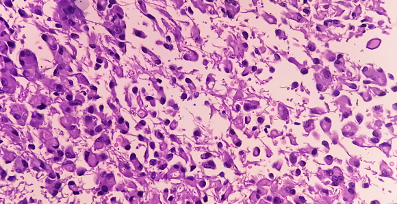
Free Images: "bestof:Macrophages in the brain before and after in vitro infecti.jpg Electron micrograph of macrophages in the brain before and after in vitro infection by HIV-I The"
Macrophages in the brain before and after in vitro infecti.jpg
Stereocilia_of_frog_inner_ear.01.jpg
Terms of Use   Search of the Day