Click Here for More Images from iStock- 15% off with coupon 15FREEIMAGES 
Set of Petri dishes with different culture samples on white background, top view
Hand with Petri dish or culture media with bacteria on white background with clipping, Test various germs, virus, Coronavirus, COVID-19, Microbial population count, Food science.
View of e.coli bacterium growing in petri dish seen through magnifying glass
Close-up shot of a petri dish for checking an antibiotic in the laboratory.
Backgrounds of Characteristics and Different shaped Colony of Bacteria and Mold growing on agar plates from Soil samples for education in Microbiology laboratory.
Research of herbs, plants, greenery, plant particles, grass, organic matter, biology, microbiology, photosynthesis, green energy, essential oils, organic cosmetics, natural cosmetics, biofuels, algae. Natural green laboratory. Experiments. Petri dishes with green plants on a light background.
Escherichia Coli (E. Coli.)
Malt Extract Agar in Petri dish using for growth media to isolate and cultivate yeasts, molds and fungal testing from Neil scraping samples. Neil scraping for fungus culture.
stx2a is a subtype of Shiga toxin-producing E. coli (STEC) linked to Hemolytic Uremic Syndrome (HUS), a severe condition impacting kidney function and blood clotting. STEC poses a significant risk of foodborne illness and can spread within communities including family members.
Top view of petri dish and culture media with bacteria on white background, solid media, nutrient agar, Test various germs, virus, Coronavirus, Corona, COVID-19, Microbial population count.
Hand Holds Petri Dish with Bacteria Culture
E. coli bacteria growing in laboratory dish inspected in a food laboratory
Set of Petri dishes with different bacteria culture on white background
Petri dish with bacteria colonies, isolated on white
Close-up shot of a petri dish for checking an antibiotic in the laboratory.
Bacteria culture growth on hichrome uti agar media plate in microbiology laboratory, Proteus colonies. Bacteria culture growth on a petri dish. Gram negative bacteria colonies
In vitro bacterial culture
Bacteria Aggregatibacter, 3D illustration. Aggregatibacter aphrophilus and A. actinomycetemcomitans, Gram-negative bacteria, part of the normal flora of the mouth and throat, also cause endocarditis
Escherichia Coli (E. Coli.)
Freshwater Closterium algae (unicellular charophyte green algae) - optical microscope x200 magnification
Petri dish with generic medium with mould and bacteria colonies on white background.
fungal culture on PDA media, this culture is usually carried out in the laboratory, the fungus is propagated and identified and used for research
Close-up shot of a petri dish for checking an antibiotic in the laboratory.
Fungal culture of Trichoderma viride
Close-up o fungal cultures in petri dishes at the laboratory - microbiology concepts
Colony of bacteria close-up 3D rendering illustration on blue background. Microbiology, medical, biology, bacteriology, science, medicine, infection, disease concepts.
Bacteria pathogen - 3d rendered microbiology image. Illustrate of Lactobacillus Bulgaricus Bacteria, Microbacterium Tuberculosis Bacteria (MTB). Medical research, health-care concept. SEM (TEM) scanning view
A scientist holding a petri dish with germs in the shape of a paragraph symbol.(series)
Liver Fluke Section under light microscope with white background
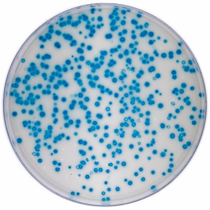
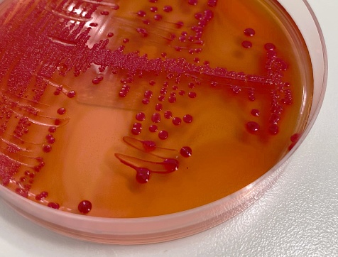
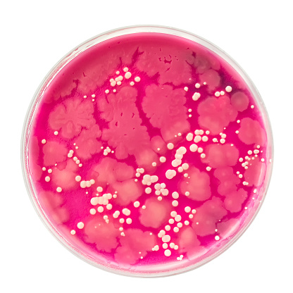
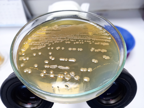
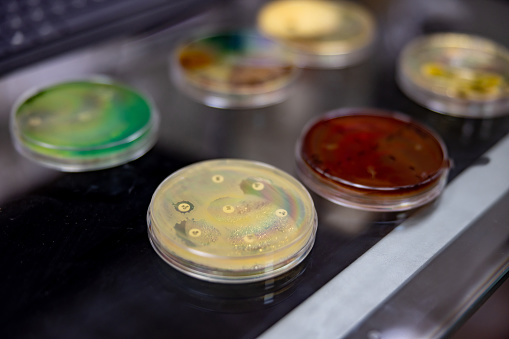
Free Images: "bestof:Macconkey e coli.jpg en MacConkey's agar with lactose fermenting E coli colonies own Microrao JJMMC Davangere Karnataka India Escherichia coli Petri dishes"
koli-bacteria-escherichia-coli-123081.jpg
bacteria-disease-escherichia-coli-62993.jpg
E. coli 2011 Map.svg
Konjugation-ecoli.svg
MinCDE System.svg
Konjugation-hfr-ecoli.svg
MinCDE System2.svg
Flagellum base diagram en.svg
Flagellum base diagram-fr.svg
QWeibull cdf.svg
QWeibull pdf.svg
Gherardi_-_Coli_-_Sophonisba.jpg
Map of comune of Coli (province of Piacenza, region Emilia-Romagna, Italy).svg
Mangalore.svg
Notable mutations.svg
Macconkey e coli.jpg
Macconkey LF NLF.jpg
Klebsiella pneumoniae mucoid.jpg
Vibrio cholerae on TCBS agar.jpg
Lactose non lactose fermenters on CLED agar.jpg
MacConkey agar with LF and LF colonies.jpg
Indole test positive.jpg
Ecoli colonies.png
S aureus blood agar.jpg
Fluorescent.jpg
TSI agar S typhi.jpg
Bacillus sps various types blood agar.jpg
Ecoli_O157_colonies.png
Simple autoclave.jpg
VDRL slide.jpg
S pneumoniae inulin fermentation.jpg
EColiCRIS051-Fig2.jpg
Trichinella spiralis muscle.jpg
Urine pus cells.jpg
Antibiotic disk diffusion.jpg
Penicillium sps.jpg
S aureus mannitol fermentation.jpg
Microsporum gypseum macroconidia.jpg
Etest Vancomycin S aureus.jpg
M tuberculosis sputum.jpg
Stool iodine and saline wet mount.jpg
E.coli operon regulation network.jpg
ELISA wells.jpg
Konjugation-ecoli.png
Asparaginase2 Ecoli 3ECA.png
Sterione bild1.png
ExonucleaseIIIfromEColi.png
OGDH-Ecoli-2JGD.png
Diverse e Coli.png
E.coli image.jpg
Ciclo celular de Escherichia coli.jpg
E coli beta clamp 1MMI.png
Isocitrate dehydrogenase of Escherichia coli Complex.jpg
Carboxytransferase 2F9Y E-Coli.png
Tsh-associated self-cleaving domain (Escherichia coli) and similar.jpg
EColiUSDAfromPinaFratamico.jpg
Sterione bild2.png
BCCP.png
ClC-EC 3NMO.jpg
EcKARI p-domain NADPH.png
Primase 3B39.png
Crystal structure.jpg
Biotin carboxylase rainbow.png
Porcine Mitochondrial IDH 1lwd.jpg
Coca-cola pl.jpg
SutterlinAlsace1942.jpg
FRAD007_061_Le_diplôme_de_mérite_d’une_écolière_de_Melle_(Deux-Sèvres),_item_1.jpg
Cucumber kosambri.jpg
IMP allosteric binding site.JPG
Blue white assay Ecoli.jpg
Trollope Thomas.JPG
Mary_McLeod_Bethune_with_a_Line_of_Girls_from_the_School_WDL4013.png
Muller Hinton agar with MRSA.jpg
Figure-1.JPG
Romanian_Schools_for_Aromanians_and_Meglenoromanians.JPG
EC 1.5.1.20 - Escherichia coli methylenetetrahydrofolate reductase - PDB id 1b5t.jpg
Echerichia_Coli.jpg
Isocitrate_dehydrogenase_(coli).jpg
E_coli_metabolic_network.png
Dihydrolipoamide_succinyltransferase_(Escehrichia_coli).jpg
Jorge coli.jpg
Metabolic_Network_Model_for_Escherichia_coli.jpg
1jyn.jpg
Coli3.jpg
Entamoeba coli Stages.png
Entamoeba_coli_01.jpg
Peptidoglycan E coli.png
Coli_and_Nungesser.jpg
Shiga toxin type 2 (Stx2) from Escherichia coli O157-H7 PDB 1r4p.png
Paul_Tarascon,_François_Coli,_1925.jpg
Galactose-1-phosphate uridylyltransferase 1GUP.png
Ecoli_O157_trans_elect_micrograph.jpg
Threepol.gif
PDB_1gvh.jpg
GCL Ecoli 1V4G.png
EcDHFR raytraced.png
Ecoli metapopulation.jpg
Obraz 34235721.jpg
WT GASP on chip.jpg
DNAPolymeraseI.jpg
Terms of Use   Search of the Day