Click Here for More Images from iStock- 15% off with coupon 15FREEIMAGES 
Fatty liver disease and hepatic steatosis body part as a medical health care concept of the digestive system anatomy and vital organ for digestion functions in a 3D illustration style.
Metastatic neuroendocrine tumor. Site: Liver.  Neuroendocrine tumors are cancers that begin in specialized cells called neuroendocrine cells. Neuroendocrine cells have traits similar to those of nerve cells and hormone-producing cells. Neuroendocrine tumors are rare and can occur anywhere in the body.
The illustration of liver is on the man's body against gray background. A men with hepatitis and fatty liver problem.
3D representation of the human body
Liver disease progression in Hepatitis B and C viral infection, 3D illustration
PAPS Smear study of a young women under microscopy showing Atrophic changes in uterus which is unusual at young age.
Acute ordinary hepatitis liver biopsy under light microscopy
Histological Pancreas human, Liver human, Vermiform appendix human and Kidney Human under the microscope for education.
Cardiac amyloidosis is a condition where faulty proteins build up in your heart. You can inherit this condition, or it can develop on its own (usually later in life). As faulty proteins accumulate, your heart struggles to pump, ultimately leading to heart failure and death.
Hepatic steatosis is a disorder characterized by the accumulation of fat inside the liver cells. 3D illustration
Captured by a scientific microscope and Canon 5D Mark IV
Intestinal tract with holographic effect, 3d rendering. 3D illustration.
Photomicrograph of Invasive squamous cell carcinoma of the nipple, Grade-II
Chronic aggressive hepatitis human pathological sample under microscopy
Pancreatic damage can contribute to the growth of cancer cells in the pancreas. Pancreatic cancer is a complex disease, and while the exact causes are not always clear, several factors, including pancreatic damage, can increase the risk of cancer development.Pancreatic cancer is a disease in which malignant (cancer) cells form in the tissues of the pancreas. Smoking and health history can affect the risk of pancreatic cancer. Signs and symptoms of pancreatic cancer include jaundice, pain, and weight loss. Pancreatic cancer is difficult to diagnose early.
Squamous cell carcinoma of the lung is a type of lung cancer. It occurs when abnormal lung cells multiply out of control and form a tumor.
Hepatocellular carcinoma (HCC or HCC) is the most common malignant tumour originating in the liver and is one of the leading causes of cancer-related death worldwide. It most commonly develops in people who suffer from chronic liver diseases, such as cirrhosis caused by hepatitis B or C. Tumour cells form trabeculae, cords, and nests separated by connective tissue septa
\
Breast Cross Section of Fat .
Histological section of kidney tissue taken with microscope. Nuclei stain blue and cell cytoplasm is pink. Scattered red blood cells. Mostly tubules with two glomeruli present. Hematoxylin and Eosin (H&E) stain. More tissue sections:
Photomicrograph of Stomach Adenocarcinoma. Gastric Adenocarcinoma.
Germ cell tumor of testicle under light microscopy zoom in different ranges
Gastrointestinal tract model and products to help digestion on white background, top view
Metastatic melanoma. Site: Neck. Metastatic melanoma is a disease that occurs when the cancerous cells from the original tumor (primary tumor) get loose, spread by traveling through the lymph or blood circulation, and start a new tumor (metastatic tumor) somewhere else.
Characteristics of fungi living in wood as a group, are polyphyletic under the microscope for education.
\
Metastatic plasmacytoma, metastatic undifferentiated sarcoma. Microscopically show cellular material of mostly plasmacytiod cells. atypical mitoses are present. plasma cell tumor.
Microscopic image of ciliated epithelium
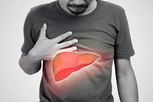
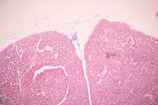
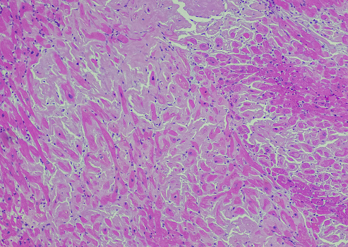
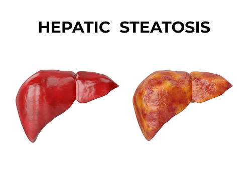
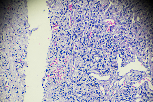
Free Images: "bestof:Liver steatosis fatty change.jpg Histological section of a murine liver showing severe steatosis The clear vacuoles would have contained lipid in the living"
Secondary tumor deposits in the liver from a primary cancer of the pancreas.jpg
Steatosis.svg
Liver_steatosis_fatty_change.jpg
Stage_of_liver_damage.JPG
Gray1091.png
Apoptosis_mouseliver.jpg
Apoptosis_multi_mouseliver.jpg
Apoptosis_stained.jpg
Gray1092.png
FMIB_52751_Sigarelus_bevigatus,_showing_extensive_development_of_the_propodium_(pr)_and_metapodium_(met)_in_a_mollusk_living_in_sand_(the.jpeg
FMIB_48602_Sigaretus_laevigatus_Lam,_showing_excessive_development_of_the_propodium_(pr)_and_metapodium_(met)_in_a_mollusc_living_in_sand.jpeg
Schematic mollusc.jpg
Gastrointestinal Stromal Tumor (GIST) of Stomach.jpg
TheFamilyDoctorNov16_1889page187.png
ChronicBackache1041.png
Radiolaria_(Challenger)_Plate_101.jpg
Catherine_Willoughby,_Duchess_of_Suffolk,_daughter_of_Catherine_of_Aragon's_close_companion,_Maria_de_Salinas.jpg
Terms of Use   Search of the Day