Click Here for More Images from iStock- 15% off with coupon 15FREEIMAGES 
Motor Neuron cells under the microscope
Cell Membrane Phospholipid Structure,Phospholipid bilayer of the cell membrane,Lipid bilayer
Host cells with spores (mold) are inside wood under the microscope for education.
Leaf of Nerium indicum T.S. under light microscopy
Epidermis. Electron microscope micrograph showing a keratinocyte of spinous layer. The epithelial cell has a polygonal shape, central nucleus with nucleolus, cytoplasm full of keratin filament bundles, and numerous dark desmosomes crossing the intercellular spaces.
Photomicrograph of flatworm,
Isolated lipid bilayer coating mesoporous silica nanoparticle 3d rendering in the background
Mesenchymal dysplasia. Site: Placenta
Internal structure of an animal cell, 3d rendering. Section view. Computer digital drawing.
Particles, medical concept
Leiden leaf c.s. under microscope
plant Flower bud of Lilium brownii var.viridulum T.S.\nunder light mircoscope with white background
Dicot plants stem show plant vascular tissue under light microscope view for botany education.
Natural agate surfaces, suitable for backgrounds and wallpapers.
microscopic view of plant cells for botanic education and analysis
Diffuse large B-cell lymphoma, or DLBCL, is a cancer that starts in white blood cells called lymphocytes.
liposome carries nanomedicine into lipid bilayer membrane 3d rendering
Microscopic image of dog stomach cardiac region
Illustration of animal cell anatomy
Captured by a scientific microscope and Canon 5D Mark IV
Plant tissue Structure, section (tissue) of stem plant tissue under a light microscope.
Tilia Stem C.S.under light microscopy
This is a 3D rendering of the interior of a mitochondrion on a white background
Blastomycosis is a fungal infection caused by inhaling Blastomyces dermatitidis spores. If it involves only the lungs, it is called pulmonary blastomycosis. Only about half of people with the disease have symptoms, which can include fever, cough, night sweats, muscle pains, weight loss, chest pain, and feeling tired.
liposome RNA transfer into cell's membrane 3d rendering
Curly marble stone with fashion veins.
Dirty pore, main trouble of blackhead and acne. Skin treatment, large pores 3D rendering
Green and blue paper clips and one yellow paper clip on a blue background.
Blue colored stem cells of a lentil plant under the microscope.
plant rape flower cross section under light mircoscope with white background
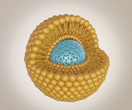
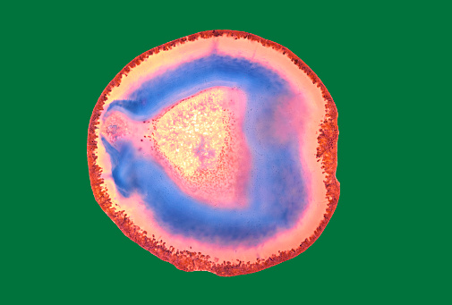
Free Images: "bestof:Liposome.JPG biology en Liposome is a small aqueous compartment which is surrounded by a lipid bilayer Own Tinastella 2008-11-11 Phospholipids Micelles Cell"
Lipid bilayer fusion.svg
Supported bilayer.svg
Bilayer hydration profile.svg
Black lipid membrane.svg
Lipid vesicle vs soap bubble.svg
Lipid unsaturation effect.svg
Lipid unsaturation effect es.svg
Pore schematic.svg
Cell membrane detailed diagram en.svg
Beta oksydacja.svg
Lipid vesicles.svg
Phospholipids aqueous solution structures ES.svg
Micelle.svg
Bilayer scheme.svg
Phospholipids aqueous solution structures-hr.svg
Cell membrane detailed diagram blank.svg
Phospholipids aqueous solution structures.svg
Lipid unsaturation effect pt.svg
Organization levels mouse.svg
Camptothecin 200.svg
Animal mitochondrion diagram en (edit).svg
Plant cell wall diagram-en.svg
Average prokaryote cell numbered.svg
Pinozytose.svg
Cell membrane detailed diagram id.svg
Frap diagram.svg
Tipica cellula procariote.svg
Stipitococcus capense.svg
Timeline evolution of life.svg
Semnalizare engl.svg
Scheme facilitated diffusion in cell membrane-en.svg
Beta oxidation (zh-cn).svg
Plant cell structure-en.svg
Scheme simple diffusion in cell membrane-en.svg
Flagellum base diagram keys.svg
F-Z DNA base pair.svg
Average prokaryote cell- en.svg
Scheme simple diffusion in cell membrane-es.svg
Scheme simple diffusion in cell membrane-de.svg
Scheme secundary active transport-en.svg
Espinosa,_1645,_A_bouquet_of_flowers_in_a_conch_shell,_with_nuts_and_figs,_surrounded_by_a_wreath_of_flowers_and_fruit.jpg
Scheme sodium-potassium pump-en-2.svg
ShockleyQueisserJSC.svg
Biological classification L Pengo.svg
Scheme sodium-potassium pump-en.svg
Scheme sodium-potassium pump-pl.svg
TRNA ribosomes diagram pl.svg
Animal Cell.svg
Balkans Animation 1800-2008.gif
Liposome.JPG
Tina Liposome.JPG
Liposome3*.png
Liposome cross section.png
Mixed lipid corral.jpg
Membrane proteins.JPG
Lipid bilayer expansion hypothesis of anesthetic effect.png
Sedimented red blood cells.jpg
Annular Gap Junction Vesicle.jpg
Bilayer AFM schematic.png
NAO str.jpg
Apotosis.jpg
Membrane fusion via stalk formation.jpg
Fluidní mozaikový model buněčné membrány.tif
Liposome Structure.png
Liposome Formation-Sonication.png
Lipid bilayer fluid.JPG
MicelleColor.png
P-lipid miguelferig.jpg
Peptidoglycan-membrane.png
Galactolipids.jpg
Sphingolipids1.jpg
Sphingolipids2.jpg
Lipid bilayer section.gif
TN3.jpg
Tranverse Diffusion.JPG
Adenylyl cyclase.png
G protein1.png
Lateral Diffusion.JPG
Detergentmicelle.jpg
Celcultuuroven.JPG
Induced fit.JPG
Antiporter.jpg
Celkweekverversing.jpg
Smalltropicalwaterfall.jpg
Cell membrane detailed diagram-el.png
Sambandhapuram.jpg
Cardiolipin table.jpg
NAOandCL.jpg
Eukaryotic pathway.jpg
Proton trap.jpg
Feedbackims.jpg
Adipos-iWatCellNumber.jpg
Combined SCLC-SqCC.jpg
PDB-7TM3.png
Hydroxylated Lecithin Synthesis.png
Single Cell Genome Sequencing (MDA).JPG
Inverted micelle1.png
Wikiresponsecurves1.jpg
Skotheimsystem.jpg
CGDB CGDef.png
Terms of Use   Search of the Day