Click Here for More Images from iStock- 15% off with coupon 15FREEIMAGES 
Supraspinatus muscle isolated within the shoulder area of human skeletal system in a rear posterior view on a white background.
Evergreen shrub, Hebe, with blooming flowers in Pembrokeshire, Wales.
Banded Demoiselle on a leaf
Acanthocinus aedilis - long horned beetle - Siberian Timberman
Anatomy of the pelvis bones, including the ilium, ischium, sacrum, and pubis, photorealistic 3D illustration. Front view. Perfect for educational or medical purposes.
Toothwort, a wildflower, in a swamp in Connecticut. Also known as crinkleroot.
The sacrotuberous ligament (STL) is a stabiliser of the sacroiliac joint and connects the bony pelvis to the vertebral column 3d illustration
Wild garlic flower with blurred background
Mediastinal view of a hamartoma tumor on the left lung. Medical themes
The pale blue males, showing off their white legs to each other, are a familiar sight along rivers and calm streams in large parts of Europe. \nField characteristics: Tot 35-37mm, Ab 27-31mm, Hw 19-23mm.\nDistribution: occurs in a great part of Europe, but are absent in northern England, Ireland and northern Scandinavia.\nHabitat: Characteristic of floodplains, dominant on oxbows, rivers and open stretches of streams. Also lakes and a wide range of man-made habitats.\nFlight Season: From the start of May to the end of September.\n\n\nThis Picture is made during a Vacation in Bulgaria in May 2018.
3D Rendering of male pelvis, hip, leg bones and ligaments labeled on a white background.  Posterior view.
Common Darter Dragonfly
Xray of ones manhood.
Hyoid bone in red color with body 3D rendering illustration isolated on white with copy space. Human skeleton anatomy, medical diagram, osteology, skeletal system, science, biology concepts.
3D Illustration Concept of Human Muscular System Torso Muscles Pectoral Muscles Anatomy
Cockroach on white
Chronic glomerulonephritis, gross pathology, scientifically accurate 3D illustration showing pale contracted coarsely granular kidneys
A Broad-bodied Chaser Dragonfly at rest in sunlight with wings open.
Yucca.  . It grows to a height of 3 m (10 ft) and is an evergreen shrub prized in horticulture for its architectural qualities
Digital medical illustration: Posterior (back) perspective 45 degree rotation (Posterior oblique 45) x-ray view of human pelvis. Featuring:
The posterior sacroiliac ligament is a compound ligament composed of three distinct bands 3d illustration
Dragonfly on dry branch.
Wisteria flowers are in the botanical garden, North China
Sage flowers
Dorsal close up of northern dune tiger beetle, Cicindela hydrida on a sandy soil
White Garlic flowers
3D Rendering of male pelvis, hip, leg bones and ligaments isolated on a black background.  Posterior view.
White flowers garden background
Die winged insect upside down photo isolate on white background top view
Field characters: Tot 56-64mm, Ab 43-54mm, Hw 37-42mm. Distinctly smaller than most Aeshna species.\n\nThe commonest small hawker. Numerous in much of our area, and although it can be on the wing during most months in the Mediterranean, further north it is especially associated with late summer and autumn, when it may appear in massive migrations. It is usually identified by its size, relative dull colours and the diagnostic yellow \
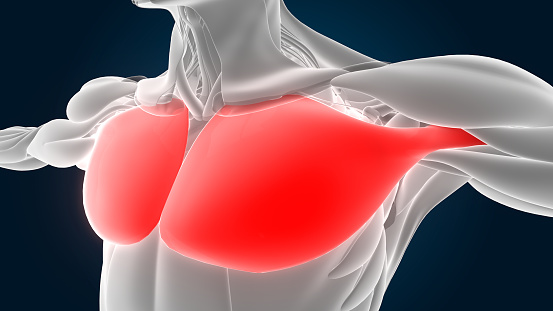
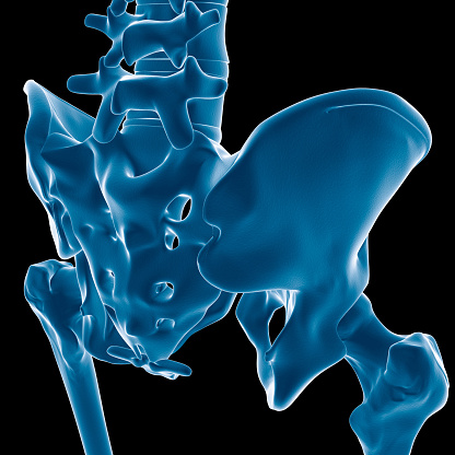
Free Images: "bestof:Lampris guttatus (Brunnich), showing the enlarged infraclavicle.jpeg Shoulder-girdle of the Opah Lampris guttatus Brunnich showing the enlarged infraclavicle"
FMIB_51901_Shoulder-girdle_of_the_Opah,_Lampris_guttatus_(Brunnich),_showing_the_enlarged_infraclavicle.jpeg
FMIB_49712_Mouth_of_a_Lamprey,_showing_the_sucker_and_teeth.jpeg
FMIB_52950_Ensis_directus,_showing_extended_animal-_1,_food;_2,_siphons;_3,_papillae,_enlarged.jpeg
FMIB_47018_Head_of_Mordacia_mordax,_showing_the_single_nostril,_and_seven_branchial_openings.jpeg
FMIB_49860_Teredo_navalis_from_Port_Costa_-.jpeg
FMIB_47676_Deep-Sea_Crustacea_with_Abnormally_Large_Eyes.jpeg
FMIB_40645_Conger_Eel_-_Embryo_Freed_from_its_membrane,_showing_continued_constriction_of_anterior_end_of_yolk_and_general_diminuition_of.jpeg
The_Quarterly_journal_of_the_Geological_Society_of_London_(13859922003).jpg
FMIB_47700_Front_part_of_body_of_a_prosn_(Spirontocaris_polaris)_from_above,_showing_on_the_right_side_a_swelling_of_the_carapace_caused_by.jpeg
University_of_Virginia_1856_(Bohn)_Pavillon_IV_+_Hotel_B.jpg
Atlas_Van_der_Hagen-KW1049B12_031-COMITATVVM_BOLONIAE_et_GVINES_DESCRIPTIO.jpeg
Pavilion_IV_UVa_Sachse_1856.jpg
The_Quarterly_journal_of_the_Geological_Society_of_London_(12645102823).jpg
Atlas_Van_der_Hagen-KW1049B10_010-DIOECESIS_BERGENSIS_TABULA.jpeg
The_Quarterly_journal_of_the_Geological_Society_of_London_(14072748922).jpg
Terms of Use   Search of the Day