Click Here for More Images from iStock
-
15% off with coupon 15FREEIMAGES
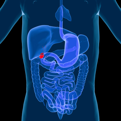
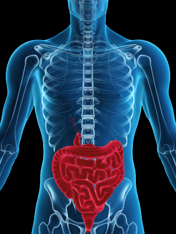
Free Images: "bestof:LUQlabled.PNG of the abdominal left upper quadrant with its contents being Stomach Spleen Left lobe of liver Body of pancreas Left kidney and adrenal gland"
Terms of Use
Search of the Day


![Image_from_page_42_of_"Select_aspects_of_the_life_history_and_ecology_of_the_Montana_arctic_grayling_(Thymallus_arcticus_montanus)_[Milner]_in_the_Upper_Big_Hole_River_drainage,_Montana,_June_15_to_August_31,_1990_:_final_report_"_(1990).jpg](data:image/jpeg;base64,/9j/4AAQSkZJRgABAQEASABIAAD/2wBDAAgGBgcGBQgHBwcJCQgKDBQNDAsLDBkSEw8UHRofHh0aHBwgJC4nICIsIxwcKDcpLDAxNDQ0Hyc5PTgyPC4zNDL/wAALCAEAAZ0BAREA/8QAGwABAAMAAwEAAAAAAAAAAAAAAAUGBwIDBAH/xABTEAABAwMDAgMFBAUHCAcGBwABAgMEAAURBhIhMUETIlEHFBUyYRZCcYEjJFKh0TORkrGywdIlJjRTYnKiwhc2Q2NzgoNUk6Oz0+E1RlVkhIWU/9oACAEBAAA/AJhDUJu52G+Wxpl1Uu64kzHFkTisrUC2UDjYOAeegrtjMRRaLPqNt9f2lkXdLbqvFO9eXilbRTn5QgdMcYq/XCffjqI2+1t28sojJeWqUVg5KlDA2/hXoCtTJSnKLStX3sKcSP6jXwu6owQI1oz2Pjuf4a4eLqvcn9Vs4GfMfHcOB/Rol7Vew7odnKs8ASHMEf0KCTqsAZtlpz3/AF1f/wBOvgmaq282i2Z+k9WP/l19MzVP/wCj204H/t6uf/h0RO1OpBK7JASodE/ECc//AA6+JuWpfv6fjDr8twB/5K+m66hAH+baSe/6+jH9VfBdtRY82mU/+W4I/hXz4xqHGfssr8Pf2819N4vwTn7MOE+nvrWaKvV7TgfZiSokc7ZbWB/Oa4m+3oEf5qTcd/1lj/FX03+7JHm0rcev3XmDx/Tr6nUFy4K9L3NKe+HGSR+W+uJ1Fck4P2VupBOMhbPH1xvr79pZmD/mxeMjtta/x1zTqGWpe37O3YYSVElLeOnT5+tfPtI+QCNO3k/+kgf89fRqOSVADTt3wTgkttjH/HT7SPZI+z94yP8AuUf4qDUrm3zWG8pPp7uk/wBSqK1OE5zZbzgHHEQnPP418OqAEFRsl7AHb3Mk/uNcRq5k9bPek845gLrmdUsDI+F3jOM49wXz+6vh1dFBwbbeR/8A17n8K+DV8Q5/ydeMDv8ADnf4V9+1kQAH3C7YJx/+Hu/wr79rYOcGJdQM4ybc9/hr59r7aF7VMXJJxnJt72P7Nc/tZbCFnE3yYyPcXs8+nl5rgrWNoQQFmYkHoTBeA/s1wGtrIc4cl8f/ALJ7/DXZ9srLt3F6QE+piOgf2aHWVkSSDJdyDg4jOnB/o0Os7EkJKpiwFdCY7n+GuQ1fYikH39OCcfya+v8ANXwaysBSFfEWwD6oUP7qL1lp9v5rk30zwlR/urida6d4/wAqsnPIACsn91fDrfTYIBu8cE9uf4V2nV+ngrabvECsZwV81wGtNN5x8Yig/VWK+DW2mQnJvcNP+85j+uuZ1lpsJyb3BAxn+WFDrLTY6323j8X0/wAa7m9N2Vq6/FG7XERP5PvCWgF5PU59a4t6WsjN4Vd27XFRcFEkyEt4Vk9T+P1rgyknWsxWePh7Ix/53KnKUpSlKUpSlKUpSlKUpSlKUpSlKUpSlKUpXzApgelNqfQfzV82I/ZT/NXzw0E52Jz64oW0H7ic/hXFTDSjlTaCfqkV892ZPVlv+gKyD20qaYl2cIbaGW3SfID3TWy0qEYcV9r5rRxsEJlQ8ozne5nmpulKUpSlKVWZYu83UsiJEuwhR48dpzYmOlwrUpS85Kug8ormq0ai3Eo1RhOeAYDZx+ea7E2y/wDiKKtQo2E8JEFPH55rl8Pv4QAm/MlWOSqCP3YVXX8N1KBxqKOTj71vH9y6It2pgFhd/iK/YIt+Mfj5+fyxXJUPUyQNl4t6jznfBV+7DlcfdtUlGPilrC+OfclkfU/yn4Vyaj6mTt8W4WxYAO7ERac+n3+K+OM6oVtLc21I8vmSYzivN6g7xxXWWdX5AE6y49TFcz/NvrsDWqkske92hTmeD7u4B/ariW9X/ckWT65Zd/xVyUnVeAEO2cnHJU26MH8M80bGrNw8U2UjuUh2hXqzKtrVlI7Zcd/hXJK9T+GnczafEz5sOOYI+nFcPE1aHP8ARrKUf+M6D/Zr6XtV+GcQrQXMjH6y5jH9CuBkav7W6zfiZbn+Cufj6s2Z9wtG7097c/wVxMrVva12j/8A2r/+nXMytTBSx8KtqhxtImqGfXP6OuPvuqk9bJblf7twV/e3X0TtS7k5scLbjnFwOR/wVzTP1CN+6xx+MbcThz6/crqFy1P4mDp2Ns9RcRkf8FdtvvU6Re3rXPtqIq0RxIStEgOBSSopx0GDxU7SlKUpSlKy/wBqhHvtt3RUOnw18qSTjkehFahSoVgj7WzBxkQmc8nPzudulTVKUpSlKUqDiknWVy6YEKP+fmcro1Jfpdpl2+NEahqXKDpKpb5aSkITuPIBqmse1C9ydPNXaPp6HJLsksNx2JpU4sAKO4DZ0O049a6bp7Wbva7K1dFWCG6266poNNTipaMEDcryYAycfjxWhzNQ221xozl2mx4S3294S4vHYZx+Ga+SdVWGGGjJu8NoPN+K2VOgBSP2gfSvs/U9ktbjCJ10isKfTubC3B5k+v4fWuVx1JZrQplNxukSKp8ZbDroG4eo+n1r0quUJAcKpTIDbQeWSseVs5wo/Tg8/SuxMphUQSg6gxyjxA7u8u3Gc59MV4mtQ2V4LU1doKw2krWUvpO1I6k88Dmu1i+WqXHffj3GI6yx/KuIeSUt/wC8QeK4xr7apsZUmLcYr7CFBCnG3UlIUTgAnPBr0PT4kULMiSyyGwFLLiwnaCcAnPTJr5EucKfv90mR5Gz5vBcC8fjivZUZeboLNBEoxnpOXENBtnG4lRCR1IHUivANRTwCpzTVybQBkqW4wAB6n9JXmhaweuMMyomnLq8zuKUqT4WF47pyvkfXvUzabq1drUzcUNrZbcBO13AUnBIIOCR1HrXq97j+CHvHa8I8b942/wA9diHW3Ub0LSpP7STkVy3p55HHXnpRKkqGUkH8K+gg9CK4qWlKSoqASOpJr6CFDIwR9KhEkfbhwdzbU5/94anaUpSlKUpWT+1wuC4WzYlRHhL+X8RWsUqGYbA1XOcJGTDZAGewU52/OpmlKUpSlKVBQk/55XVXrEjf1uVW/aVZnby7aM2B+8RWC8t1pl1KFJJSAkgk889qow07qEQoMhOhSq7Rn1BsrU0lnwPPhBSkj9oc14bjp+/G1uxIOhH2npMlS5Ti0trSUbgQEAHjBH4Y+tafqRExm425TMS6lpEFxsuW9pDikqJRhJCgR2NQsLTF7+DCIhbsFxFnLfLKHN697h2EngKwRnHrXZbI0rTgfTK09Lnpn2+M2wGWQ54akNbFNLz8ozzk8cmuq0QZukCpN2s0u8OSYDDTS4zIe2KQkhTRP3Rk9eleaJpG9NMsNyGlPIhQGVuQf+ylEOOK8Er77QoYHTOM1qUNxuTAZcSwpptxsEMuI2lII+Up7fhVKm25StNarbjWzY+uUtDWI/K0kI5AxyOv81RF105eES7n7xHTcGi3DeUmLEDKXm0PKUtraDhSsYPXnpXK4RnbzNn3ayWaSxDaiNIW0tgsKlOIeSvyoIBJSlJGe+cVP2aS3qTUt0nfD3DbPdGY4MuOUeI4lSlEbVDnG4dute/Q8CPE0xEW3BRFecSfFHhBCjhSsbqs9VvWUlMS0Rn3EuFtE6OpexJUQkOAk4AJxxVemajt2opvhTn34tlaXgsLjuhyaoftAJ4bHp97vxUldNfWa2RQ3A3PyiQ20wGVoSk9MqO3ypHeod9tx72cWVUxtx2GJaHbgllCvMzvUT5epTnaSPSoWOpFxXOh2+HAchruEMtFEZ1MVRO/OUH6AZI4Jr1Mx7lFtV408zESiXcLqWNkPLTLbPhJUsoJ+XKcjPqa7ocK4rdh21zx4SpTxt11RysuIbb3IWlfTKkYSVD19RVhs94s1n1BeLUuSxFX70y2wyTgqHhISAPzGKgoPvb7unPcJK0S2vf320KzteSHfkV9CCeexArySXmZMOzPXlosWpb05SmphKWhILhLYex2xu+lWH2XSxJsdxbbCRHYuDqGA2tSmwjCThBUAduScVYEJA1q6onk25AA+niKqbrwyrpAgrCJc2Owsp3BLroSSOmee1dX2gsxGRdoGMZz7wj+NeyPJZlspejuodaWMpW2oKSfwIrvpXldnxGHfCelMNuYztW4EnHrg1x+KQOf16Nx/wB8n+NehtxDqAtCkqQoZCknIIrsrMPanHD0y2koQSG1jzKx3H1rts7t0+08Vlu9zLjKadWq7bFAw2UYOEJ44UDt4HPXNeew3G8qu0O6XZyd7hcZziIpROSWwCVeGlTQT0wPU1aJdwuMPVclES0LnoVDZJU28hBQdznXce9WOMtx1htbzJZcUkFTZUDtPpkda76UpSlKUqp/GIFt1tcW5r/gqdixw2VpO1WCvODj6ipRWp7KnrcWPmKep611J1fYFIKhc2MDPrn+bFPtfp/cE/FGCSrZxnr6dPrXb9p7GQ6RdYhDOPE/Sjy55Fdf2v07uKTeYQI6gugV9GrdPEZF6gY/8dNc1aosKEBarxASkgKBU+kZHr1ojVVgcICL1b1E9AJCf413LvlpQ6tpdyhpcQSFIU+kEY9RmuI1HZCSBd4HAyf1hH8a+i/2ctF0XWD4YOCv3hOM+mc1yVfLQlW03SEFehkJ/jQXy0qQFi5wik9/HTj+uuQvNrPS5Qz/AOun+NcxdbeoZE6KR9Hk/wAa5ifE/wDamP8A3gr4m4wlEhMuOSOwdT/GuXvkXPMhnj/bFDMjAgGQzz0G8c19EqNjh9nA/wBsVy8Zr/WI6Z+YV9DrZGQtJA75riQwo7z4ZPqcVzSWzgpKT6Yri4hp1BQ4lC090qAIr6hKEJ2oCQPRPFQqCFa6d82SLanj/wBRVT1Zpry2SrnquI1AmxYcv4a8Q9I5wjcNwCTwc5/LGapFnER+1R7jBsNvRbGI6m5UNx07n1gN+YDbkZOOPv8AatW9m6t+gLQ4G0NhbalBtHRGVqOPy6VbKVleu7ZLmalmvQUwkqatG6Q5IIJLW5eUpBB83ort+dVCRabexazfY9kj/Z0RwlURbuFlwkYIyneOSCU98ZHHXZtHILWi7KhSAhQhNZSDkDyjvU7WWe1lIEy2KKwnLbg5z6j+NW21aItVlcaVb3p7KG3FOBoS1ltRPXKScGvsTRVqhXRE5r3klpanWWFvqUyytXVSEHgHk16oYzq66HcTiLHAGOBy5U3SlKUpSlK+YFMD0FNo9BTan0H81cPBbznw0Z9dor54LeSfDRz18or4Y7J/7Fvj/ZFcVQ4ywAuOyrHTKAa+CFFTjbGZGOmGxxRcGK4oqcjsrUTklTYJrr+E27n9Qi89f0Kef3V9+GQQ34Yhxwj9nwk4/mxXFdotritzlviKV6qZST/VXUrT1mWrcu0wVH1MdH8K+/ALOAB8Kg4HI/V0fwr4rT1mUcqtMEn1MdH8K4K01Y17t9ogq3cnMdPP7q+L0tYHBhdlt5//AIyP4Vx+yun9xJstvJPH+jp/hQaV0+nGLLbxjp+rp/hXBWj9OLQEKscApHbwE/wr4dHacJybLCJxj+SHT0p9j9PFRPwiIM9cIwD+VcDorTZ62aL+STXIaO08kYTao6RjGEgj+o0+xun8KHwxnCsZGVc4/Ovg0Xp5OcWtrkY+ZX8a9Ns09arM647boaGFupCVqBJJA5A5NS1ZL7VExHLzF+JMhcBMFwqWpWza5u/RjftJGT2HUgA1FmDcjebMl+xWxd19y3RWW1gMvAbMrUNuScE+m3t9dI0EHBoy3pdQ224kOJUlv5QQ4rO36VZqVjHtRMYaoeVcoqlREWwFp5C0t5eyvDZUeoIydnfH0rzmNJN1KH9HsG6CGjZELyA0pAxlzPcgfePIxj8dU0eVnRtmLiQlfubWQO3lFTdZl7UHAibbgqQGj4a+C3uzyPpWm0qIig/aa5Hfx7uwNuOhyvmpelKUpSlKUpSlKUpSlKUpSlKUpSlKUpSlKyj2jqYGqoge06b4k2x4qjjGW8KyF4J7Hg/jVWt9pUxbkRI1rnSRKipV8ZMxKTHCyk7EnfgbcDaMjryK1b2doKNB2lBzlLRTknJOFHknuT3q1UrIPaE5FGq5yJNjXdUqs+9GxWfd3EleFqTnhOD8304qtPwg1JctTabyu8e6Bs3z3gEhJ820YXjoNuM9OetbPo1Kk6LsoWnaoQmgQTn7oqdrLPao6tFyt4SUgeCrqAe9anSoWF/1rux4yGI4/t1NUpSlKUpSlKUpSlKUpSlKUpSlKUpSlKUpWXe0BMtWqowtrMoy/h6ytxhwpHhBR3JUAQTk46dOpqosvWx5cFcO0X37Ne5qbmIDiwSSU7ghG7O0kjPr2rVfZ0lkaBtAjtqbY8IltCjkpTuVgH8sVaqVj/tEbm/aa4qtvvyXBa0Kl+CvyOMAuApIAJCupB6dc1W5PwJ+QtLL97b0imD4KoyGV8rKsg5252hWOpzn6Vteklb9H2ZWMZhM8f8AkFTNZl7Tt3xGByr+SV0H1rTaVB27P2svRP8AqowH8y6nKUpSlKUpSlKUpSlKUpSlKUpSlKUpSlKUrKvaT7j9omFXSfPiRE25excNskl4rwkEgEgHJGOh6Gq0qbdCpifObuzF9RD2xIDbBV4mzZnCgjByCeTnbWp+z8LOhLQp1K0uKY3LS4MKCiokg/nVnpWO+0JMV3VdybnXWVBSbUhEdEZokyHSXMNlQB64+Xv+VQHxIpWb2mRc1X4QubauEkLKflOcIwSOoXjoMVs2kVb9HWZW3bmEycbcY8g7VN1lHtaCvf7YQ8lvLS+qiM8ivfbtQ6gj3uOzdJsaUtUZ2RcIMdgD3AJTuSCsE5JPGD1r022/X9CrDcrhJiPQb24ECM2zsVH3pKkYVnzcDBzXsdvyLRq+8pdt9ykJU1H2qixVOjhKsjI6datzLnjMoc2qSFpCtqxgjPYj1rtpSlKj7pd4dnjokTXFobW4GwUtqWdx6DCQTUf9tLHtKjIeSB+1FdH/AC16bHqO06iZdetUxMlDKtiyEkbT+YFS9KjbperbZkNLuMtuMl1RSgrz5iOcV5Pthp3AJu8UJIzlS8D99e613i33mMZNtltSmQooK2lZAI7V76UpSlKUpSlKUpSlKUpSlKzHXaU/bGKVPFQXbVspip2kqUpRAWQoY2p7ntmqm6+GbvBQ/rFh2aIfF3CE4hBIR5DjIyefxzzWq6CUpWhrSpUn3pSmcl8HPiHceastKxb2lyEQtVzHn5SdjlubaZZCQpSHSV4dxkbQBkbxyM14ZLmL88pnWkf4p7oFKuLiklstY5bAB2g5+7j69a13SfOj7NlwOH3JrzjoryDmpqsv9qFubn3G3lePIyrGXNvU1YrBogafbLEe7SHYq1KU+y602fG3DncrbuJ+ua42rQwt02Ct+7y5kO2kmBFdSkJZyMckcqwDgZ6VM293ff7u1knw/B69soNS9KUpSoTUSwgWvJxm4Mj+s/3VHPSntUyVwoK3GrM2SiTMQdpkH/Vtn9n1UPwFeyxxY8O93diM0lppsR0JSngABvjirBSoa6eCL3aS8knb4y0q3YSnCOSfyJqJc8XV7yozB26eRw68k8zD3Qg9m/U9+g4qQ07Gbiybw0y222ymbhCUdAA02MY7VP0pSlKUpSlKUpSlKUpSlKVl2vpF2iaqMizTIcd9FmcDomkBtSN56HOd2cY7etVK3SoQs8Zhm9QRpR2LsltuICXAsbdwAKt5BOM49OK1f2fhoaCs3gq3M+7jwz/s5OP3VZ6VkmtlXhjVV6Xa3ovhPWdCJSJAAKUfpOUHPKuvHSq/MbjNWeRCVPtSdLmKHEuBolRXvPlJCt4G8dfy6VsWlM/ZKz5UVH3JnzEYz5BUzWZe0yQhi5QQuO27lk4KyRjzfStNpUHav+s1/wD95j/5dTlKUpSqvrSA1dYdshPreSy7cWgvwl7VEYUcZHb1+le253WHpyCwy2wVOrwzDhMJ8zhA4SkdgO56AVH6SRck3G9uXZaFTHHmlKS38jY8MYQn1AHfuc1a6VVdU2lm83ayw5EhxEcreU602op8ZIR8hI+76177pd4tiYYjNM+JKe/RQ4bQwXCB0Hokdz0FePR7U5qPczcltKmrnKW94IOxJKEeUZ9BgZ+lWelKUpSlKUpSlKUpSlKUpSsj9p8ZUy+pjqnQ4DJtynVPyd2XNpVltJB4BGSR97H0qFhs3C4XuLeXbNZ0XSLABRaCg5c5AS4V9MnPB7c5rVNCrK9FWlakJQpUcFSU9EnJyP581YqViXtKYM3VV0Sl23MOxbe08kyiQt9Hmy2ntgnHHXOKiJzUj7SPXp3T1nEr3DwvgSSS9uOR4mNu0kDnOMY75ratJAjR9mBSEn3JnyjoPIKmqy72o7PicDcMnwVdx+1Wo0qEtSt1/vo8vldaHGM/ySetTdKUpSqtrWe7botrfYhuTHviDaUMN9VqKVY57D69q77LYnWJTl3uzqZF2eTtUtI8jCP9W2Ow9T1Jrr03dIl0vF/chuh5tuS20XEnKSoNpBAPfBzVmpVS1ZdHbVebIYsV6XLdL6GY7Q+dRSOVH7qR1Jr22WyKhOO3O5vJk3Z9P6Z/7jaf9W2D8qR+/qa6tJXFi6/GZMZ4PMfEnEIcT0O1KAceoyDVlpSlKUpSlKUpSlKUpSlKUrI/aSuHF1G5JnWdq7D4X4LTAWPEbKlLy4EY5AwOR0qCas8+PKbszlpszmp3ofjNz0rAaSkEBKVIxxlOeMcnntWq6DbUzoSytr2b0xEBWzpnvirJSsS9pSrczqq4OTrGbpJdgNMQylYPu6vOfEKeuAceb6VAyrFcV3WZB+CxTqdEEvrfTJHu6UkkFITnps6J6556VummGvA0raWf2IbSeufuCpesw9pjbjl2heG2pWGDnBxjzGtPpUJaW0pvt9cHzLeazz6NJqbpSlKVB38kSrIACQbgnP0HhrqMlSZGrJLlvtz7jNoaUUS5rRwp8jq00fT9pQ/AV7tPRWYNzvESM02zHZcZQ22gYCR4SasNKg7mUjU9lKjjCJJyf91NRjj7usXlxojim7C2ra9ITkKmEdUIP7Hqrv0HrXv0xGZhpujEdpLTCJ6whCBgJASgYAqwUpSlKUpSlKUpSlKUpSlKVk+vosp7WLjkK3uyVpsykPrSvaGmVKVuUDuGVDHCeh5zVPcj2NUdM+PCucjTKY256Vvy6FpKBwsufKOOceXkfhsmgkIRoKxhtCkIMNBSlRyQCM8/z1ZKVjOvUyG9VX+THtkmWk2tpiW60rZ4DJ3ErB3DJ4PlqtTkWNtMmW21efgXuodTOQ4ou+KSpIBXvIKT03dOMVu2m0qRpe0pV8whtA5/3BUrWb+0KQpm9RkpCSDHB5GfvKqx23Uz8283WFKtMiCzCaDqHn1DLycqBUAOg8vrURozXy9WSUpDVvZQpCnA2iUpTwTnAOwoA/HBOK9rGobLatSX1mdc40Z1TzR2PuhJP6JPQHtVsQsLSFJIKSMgjuK50pSlVbWVuTdWrTCW+8y27cEhZZVtUU7F5TnsCOPzr23O6QdL2tlCGck4YiQ2R5nVdkJH9/QDk14NIIniTe3LopHvrspC3EN/I2C0jCB64HGe9WulVHVFpN7vtjhuSXWoxD6n0NnBeQEp8hPoc84qVuV0h6fiR2UM5ccIZiRGQNziscJSOwHc9AK8Oi0zRDufxHYJirg6p0NklCSQkgJz2AwPxBqz0pSlKUpSlKUpSlKUpSlKVkPtHWlerCwHrjFX8LK3HohUQ82FKJZKRwM9So8+gqrvMQBNaUq2XiMw22EfBCV+I+Tt86SPKSOvTBCeTnpsehEpToSyJQlSUiIjCV9Rx0NWOlYpryU2NZ31l6RPjtptbanGo24iYnkeGcAgDJ+brziq3cV29yTM8l6ValshKbYEK8VDvm2lSSnaB1O38+tbzpsbdL2kbt2IbXOc58gqWrNfaEHlXuP4UUvARhkgZx5lVapNjkSrzc5CpARGmW9MRIR8yFZXlXp96oKy6VvbV5sz10ft4iWVhbMf3VKg4/uSEgrzwBgZwO9TlrjNvX2+rfjtqIkt7FKQCceEirABivtKUpVa1fcWrWza5K2H5DvvqUsMMpypxZQsAfTr17Vys1jfTMN5vTiJF1WCEBP8nFQfuN5/erqfwpp2ZHn3bULsZ1LraZiUFaTkbktIBH5EVY6VUNU3dNrv1kW2w5JlOJkIYjtjKnFFKcfgPUnoK9tmsTjMtd4uy0P3d5OCsfJHR/q289B6nqe9fNKyo8wXd6M4h1o3J0BxByFYCQcHvzkflVjpSlKUpSlKUpSlKUpSlKUrIfaRKtStVmDdX5jSTbRIjeD8hfQpZQFcH1/CoJc26/EkLlXyYvVAgEMPssARmm1kfMnZkYweCMkgYrWdDFZ0NZfFKi57ojcVDBJxzVipWJe0u4WpvVNxi3Ka9HkNQW34AS3lC3juT5iBnjj6YJqHn3WYi6TH0XxDmqzADLiwyj3Yt+Ykjy5HlwAeuTjpW36aChpe0hZJV7m1knGc7B6VLVnWvAo3tjb4P+jJz4uf2ldK0WlQtnO67XzzEgSkDGen6JFTVdEiUxEb8WQ82yjON7igkZ/E10C8W0nHxCJn08dP8a72JTEkK8B9p3b82xYVj8cV30qFvv8AplkG7B9/B/H9GuoyVLf1TIct9ueW1amlbJc1s8unu00R+9XboOa9mnIbECZeIsRlDMZqQ2httHASA0irDSoO4Np+1dmcIGUsyefyRUa9Jk6uech295TFkQrZIlpyFSvVDR7J7FX5D1r3aUiR4Ee4xIjSGo7M5xDbaBhKBhPAqwUpSlKUpSlKUpSlKUpSlKVj/tKuMuFqpbdukIRKftfhupdjhaQxuXuUlRPCvp3/ACqCQ8zGnm0t6wQq0KhbXLqtvLrb2QU+fdyRgDd0HStc0QEDRFlDchUhHujeHVDBWMdasNKxL2hT34WrLw3FehJMqIy1KU+1uWwxg5cQc+p6YPQHtUU7JZN1utmOpIKk+5bvjvhEPPO+Y7N+/aTtyMjjtitu06pKtN2tSSCkxGiMDH3B2qUrNfaEhSr3GISD+rDsT95VaVSoKxEm7agyAMTkjOOv6Fup2oDVcaPMt0RmUwh9pU6PlC0gg+cdjUJdoNtkzlWKx2e2++bQZEpURCm4aexIxys9k/meK9WktPW/TV4uUC3NBCBHjqcV95xZLmVH8auFKq2sbaLuqzQlvOtNOTv0imThRT4TmU57ZHGfrXunz4WmbYwyzH5OGYkNgAKcV2Skf1nsOTUfo8XIvXpd2LQlrmhZba5S2C0jCQfvYHf1q10qqakta7tf7NG97ejx/DkF5LOAp1Pk8hV1APfHNSNzucTTkBlpqMpa1Yaiw46fM4eyUjsB3PQCvHo4zvc7ibkG0ylXB1S0N8pRnBCQe+B3qzUpSlV3W0tUDScyWgyMtFtREYkOEb05CcdyOKyl65XFmdfJslWomYK2QLfGRMWpbK9pO5YBJA45z8vepnR0e5XLU0+0Xi5XkqiRkPJdTMWgLCzxkA9QB179cVPS7Q9OuBtVmut4R4ah75NVNWpLI67EZ+ZZ/mA6+lWbSinF6bieNIdkuJ3oLzxyte1ahkn8qm6VVnoEi56mnoF3uMRlhhna3HdCU7lbsnBSfQVCym7lcpjlusmorn4cYn3ucotqQ3jq2k7PMv15wn8auFgW87YLe5JdLz6o6FOOEYK1FIyak6UpSlKUpWXa0aemapuDMW6x7c43Zgpa1/M6gqX+j6gYPr1HGOtVhy7yAXbjIXa24a4qAq0uoIUs5SkEt7jzk/KCArAJrVtDjboayjCf9DbOEdBkdBVipWN67E13UOpjDuMKEiPbWlyUPDcqUjCjsAPToRuHPmxVbkylv2qXeJNvt4tCo4bbthKlBK9ysKDfUEHJ2Z4zmtTs0fUremrev4za22UxW17lQlHajYOp8T07157FftSTr7HXI9zXY33VssPCOpp17CCreAVHCcjA9etdWu3FovEfYSMxxnyg/eV61oVKg7Evdcr9wBtnAcd/0TdTlVzWDUh63Qmosj3d1c9gB0JBKRu5IB4zivW01bdLWdxWfCjNguOurJUpau5J6qUah9Lyptx1HeZkyKYgdajllhZ86W/Pgr9FHk47cVcKVVdYXP4WuzSEx3ZLpmlDTDQ8ziy0sAfQZ6k9K77JY5Dctd3vLqX7q6MAJJLcVB/7NsH96upNfdN3Rm6zr45GeDrDU0MpUnlOUto3YPfnNWKlVLUt2Ta9QWgpiuy5jrchEeO11Wo7Op6AY6k9K9lls70R926XV8SLq+nC1g+RhHUNtjskdz1PU00pcGLnGuUmM6XWjcXkpczkKwQOD6cVYaUpSqp7RTLToa4Kghwy0+GWg38xV4icVmQn+BGvqnPjarm9HbF1T4Q3MpIIP3OvHHqDzUhpOH4+ppVv0nPl/BnoSfeJslB3IO87ktHAyT0yflxx0rTn37VpKxJJAYiMgJbQkFSlqPQAdVKJ/MmvPol5cjSMB5xpTSlhaihQwU5Wrg/WrFSqZOYus7Vc+DC2xIr0dgyZwVlzb5xsQnsT+12/GpKe9a9I6c2JQlmM2nwmWUDKnFnokDqpRP8AGvVpgrVpa1FxGxfujW5PodoqXpSlKUpSlY57UkwRfX1XVERUFNuSpPirIX7xuX4eAkZPG76etfHIV5GoY6lo0+L2uAfBBQsMqaBBUrGCCfxOfStF0UXToqzeMlAc90b3BHy5x2qfpWMa8htuazlrfjQ3HVtspty31bR71gjafKd3GDg4HWo6Xb7z9o7qhu02lN7MHD0fgMBjzArHk69Oc5J7Yq62CJO1ZZ7Uu4NvQ7IzHaCYnyrmKCR5l+jeRwnv1PFT15mxm9RaftyFo95MhbgaT1S2GljOB0GSBVb9oi0Iu0TevaSx+0R941ZIOsbfN+LrLchhi2IC3XX2ygLThWSkHnHlNdNn1kLnco8KTaJtuMtkvRFyduH0jBOME4OCDg17LRLYbn3pDjzKCJvQrSD/ACaOv/3qfqs6zuMe1W6BMlZ8JFwYztBJ6nGAOSfpXVbbZNvE1q9X1ssholUK3k5DAP33OynMfknt616rXJakawvqG3UKUy1GQtKeqThZwf56sNKhbyE/ErKVY8stRGen8k5UauYvV0hyJbnXGrO0opkTWiUqkK7oaP7PZSvyHrXs01EjwXrvEiNJaYZlhCG0jASPCb4FWClQU4tp1XbFqJBTFkHJOABlvJNRb7z+s3nIkVbjFgSdr0pBKVTD3Q2eyOxV36D1qS0vHZiwpkeOx4DDU15DbY6BIOOB2H0qfpSlKq3tBDJ0ZN94lrhsbmvEkNnCmk+InKge2KyNbcdty7ZvshLZY2xZQjkC5qUFYQSU+bIxnk54xVg0ff48HUV3uM9MiOtUFlpEBUfY6pQUUjagJAUVHpgdMZq/Wq0SZs5F7vzaRLHMWJnciGk/1uHur8hXs0mtLmmoi0hISreQEjAHnVU3SqrLu0Sy3u7SZCy44pqO21HbGXHVYXhKR3JzX232iTIWu83wNrnbVGOwOUQ0EfKn1UR1V/NxUlpjP2WtWevujf8AZFS9KUpSlKUrKNdR5o1dNlw7LFuqU2TY6h4gKZBUvDicjtjkDmoFm2OWy0O2dq2xRbSyn/Kjr+SkK5Vlwp4V6fdAOOTWq6K/6kWX5P8AQ2/kGB8varBSsY1y2v7UajW3YmrikwGAqQVgLhE5HiDPQY545yn0qHn26NIg3GCLQlSWGMG9JnpLjxAKuXCACSRg4J6YHNac3fzBsVnttuYEm8SITZZi5wEDYPO4fupH7+grhHsbdoulokyXzKusuUoyZa08uHwV4SP2UDsP768utDPF3Z90bCk+AN36LdzuV3xXvuunJl3kalYcUGo1xgNR2HArJCgF5JH4qFeS127UM+82V+7wWYLFnYWkeG+HPeHVJCNwx0SBk888164GnrNcrxfJE22RJDwnAb3WgpQw0juataXG8DatOO2DURqBKHU21JAUPiDJxwehJryXe6TZcs2awqR72f8ASZZ8yIifqO6z2T+Zrhpi0MWS73WLGCijYwtbqzlbrhCypaj3J4q00qr6ttPxp+zRFvOtMGWoveEcKUnwl5TnsD0P0Ne64XGHp6Ey0hrKlYaiw2Ejc4eyUj+s9AK8OkBNxdzcVNqmmcS6Gh5E/o0YSPwGBmrRSqfqWzJvWqLPGekutRQxIL7LfHjpy35CeoT0zjr0qWut2jWKI0y0z4shY8OJDZHmcPQADskdz0Arz6QbkNWp/wB8U2Zi5jy3/DHlCyrkJ9QOmfpVipSlKqXtH92+wlxMwq91T4anQlG8lIcTkbcjP4Vkq5jaY8xUrUkdyAtpCbVHwFGKvB8NSkpOUFIwM87c81dtGsLm+0G4S7tcoV1nx4DIZkRsFDSVEghIySCSOc9c1Zbpcpl7nPWOyuKZDflm3BIyGAfuI9XCP6P417dHtMxtKW9hhJS222UAKJJyFEHOeSc1P0qsRbXCe11c7mtrfNajsNIWvnYkhRO30JrzXubLv5k2exyfCbbBTNno58LjltHYuH1+7+NTWmm0taXtTaBhKYjQA/8AKKlqUpSlKUpWQe0Zl+Vq5YiNFL8W2CQ4v3gI3NBS9ycEjOePN1T+dQ80RZlybubunrmuwojKSu2qmA7nCfm8PfznPy985rWtHONO6Ns7jDZbaVDbKEE5KRt6VO0rGdcQX7hqm+IjMvlliMy9OLcjZ4jW35AncAeh5weuO9Qstu0sMXJ52xXdOnPdUtxreqQf0a/Mdxa38eYfKckYJrUtE2eLYdLxZbrynJMiK27KlyD5lYQMAnslI4AqOTdJmodV2WcwPAsTcl1Mdah55a/CX5x6N4zj1611a8dkNXmOGVqSkxxkAkc7lelaJSoGxK3Sb7kkpE9Q5/8ADRkViht592sq7rayizynHUxHIrgTKcdOSkKxwcEcA9e9c4sGUvUligSI7ttvTk5ZZktuJLYaCnNxCMkFQ6ZPTjFbYhu1aTsy1qUiNHR5nHFfM4s9yeqlE/majdLzJdyvF5lS4ioe/wADwWVnzhvarBV6KPJx24qfuqttpmK64YWeuPunvWGMtTALIudGuUYKiBUHwJG1c1zKMeIguc9egIOOfw52a8SLVqOyLlQ7v8aWHErgJfK23UlB2EArJ5OMrOAOfStasljfalLvF4Wh+7PJ25T8kZH+rb+nqepNcdLTWLhIvr8Z3xW/iKkBYOUna2gHB7jIIqy0qm6nvXwjVNoDMZ2VMfjSG48dscrWS3jJ+6ngkk+lSVisjsRa7ldHUSbw+MOvAeVtOchtv0SP39TTSktubBnPMvpea+ISAlaenCzVgpSlKq3tAefi6QkPxGW3pCHmFNtunCFKDqcAnsKy1TtxYGrJzSrQ5cH0pTcGltlv3ZBRjydd5GPXzVJaSiTHdSz7dYrtHegPRUF65NHK2wVklKSfmXzgE/L05xWnuu2vSViBI8CIyNqUpBUpaiegHVSlE/iSa4aQcde0tAdeSUurQVKSeqcqJwfrU7SqTOTcrhqq5WuATEYcZYXKnpV50p8w2IH7R9e341MTnrXpTTSzhLEZlsobQkZUtRHAA6qUT+Zrv0wpS9K2lS0lKjDaJB6jyipelKUpSlKVjntMj2VerFv3luY4BbEtRUsEhBfUpe0LII9Pw65qEmOXZycmQ7brynUzUHLcJtai0UbsHK85xgk57EYFa/onf9iLJ4gUF+5NbgrrnaKn6ViXtDMJOp7wq4Tbgy+phlu2sR0nw33inOFKA65A4Jx3qDuCZzbt1uwZvX2jMRIfglrhtpW9PLm3oAAd3U8itKsTUrWFptypjTjFgbjtAR3E7VzVBI8yh1DYPQfe6nipq7SojGpNOQA40h9Trqm2e+xLShkDsBkVAa6aU7fmSGwrEVPfGPMutEpUHYlI8S8qSAT7+5u+pCU1iNr2Mqgzmocu4vuiUV21p5QRESOC6kBeec5OACe2K7LRc4FqnWy5yYEl9DNyPi3fKlJTnxMMhO5WTyMY9efrrFttc293FF6viFIbQd0C2qHDA7Lc9XD/ADJ/Gvdan2ntU39LawpTfu6VgHODsJx9DUndRm0TR6x3P7JrArUwWvhUlxF4nj4eXI8dK17bercgbgrO7b1J78Y6VP6KKW9SWEKjXNUpSXUO3B5Sg0+nwtyUgKPUccdOKvUqZJ1ZMct1sdU3ZmlFE2chWC8odWmj/aV+QqS0wwxETc4kVlDEZiYW22kDASAhFT9KgpQT9s7cSBlMKQdx/wB5uol2XI1k+9Ct7q2LE0otyZrZwqUR1baPZPYq/IetSWkYjMC1yYkZlDMdma+hptHRKQs1YaUpSqr7Q4rs7RM2Kw60y88ppDbjvyoUXE4J/OsmcsbjzFxhMwLSiTBQ2qTMQ5lMwY/SITlHdWMn7ueKsul7zbrRqS93N+0MWRAhstqgM8ulzeoJG0AeZXGAOowautotU25TUXzUCEpkJ5hwQcoiJPc9lOHue3QV7dIEHSsAp6FCj/xGpylVSVdodhvF4nTSoIUiMhtKE7luqIVhKUjkkmvsS1ypZcvV8P60G1mNFx5IaSP+JeOqvyFS2nTnTdsO4K/VW+QMA+UetSlKUpSlVK3ztV3FDkhn4OhjxnW0JcS7vASsp5wcdqixqrUibp4SI1rlwY8htiXJaU4kJWtYTtRn5lDOT2HStBrGvaWlpnVcuUtqbJUbP4JZiNbygKUv9IcpICRjk8H0qGefiRrk4FainhhTCXF31bJ3oXwA0f0focAZxg1r2hwBoaxhLniD3JrC/wBryjmrBSsP9oMxqHrC9rccfWpyIw0ktMhz3Q9S6cjyj6g5ymoi4yISrlcvA1e4jdF8Ry8L2kSshY8L5ceg254xnrWsqv6LXp+zwrez73c5URsRIoPbYPOs/dQO5rys2EW292aZMc97vEqSv3mYpPo0vyI/ZQOw/vrz61bffvbSGk5CI6SevdSv4VoFKgbCMG9YIwbg7jHbyp61ilpuDUp+3vWY3OPfWw8Lg/4anPGZRwQ2rBGSMYJ+WpDSk5Ny1Bp74c5cUWVNwcPu0xokOvEOK8QKxgYGOM9c+lajeb1KkTzYrDtVcCB7xJIyiEg/eV6rPZP5nivmlrVHstxu0KMVKSCytbjity3FqQSpSj3Jqbu+42acEEBXu7mCfXaawuzXeEGEP2S5zW1Lg+PfFuNBS0pG1KktjZjdwfy+tejTDa73dNN2233iZ9nUtvIS2pvY6VbCV4VtHkOcZznritcuVxt+k7Oy03HwOGYkOOnzOr7IQP7+3U149Ge/lm6u3MMImOz1LcZZJUlryIwnPcgYyatVKp+o7Mu96jt0YzTHimK8JCGyQ48jc3lIPYHjJ64/GpadcIGm7fHYbZAKsMxIjPzOK7JSP6z2ryaK98+DSDPShMozpBcQjlKSXCcA9x9astKUpVQ9pb0KPoaa9cYrkqEhbJfZbVhS0eInIFZdcLdBj2yQ67px9EJ5LYtO15Kgha0ZSXDu+bnjt61aNC2J37d3CZfrU3HvDcRtY2uhxCQVKSFJ5ODhI6/XHFW28XWbdJzlgsKtjyRibOxlMVJ7J/acI6Dt1NezR0dmJpG2R2C4Wm2doLhyo4JySfXNT9Kq0Wzxn9dXK5SEl19hlhDG85S1kKyUjsT69a6r7cZV2MmzWNzYUJUmbOTgiOMcoT6uH93U1L6YSEaWtKR0ENodc/dFS9KUpSlZ7bJsvULD1nthejxG5UhM+cBtI/Sq/Rtk9VEdVdh9anbzHhWawwYkZLcdhMyM202OM/pU8D1PWrLWSa/ny42rJ7UO8MW4Ks48cSNu11G5zyozzv8AQjpz9KgZMm2uRfFXfG16YdjBCovhJ3cHj9GCFbd3UZ3ZGela3o0tq0XZS0tS2/cmtqlAZI2jGcVPUrFdbSbk3qvU7EK4RY0N2DH9+S+kby3gjLXOVLxnjp+dQV4Ed22zLfIv0FjTbcVAYbDHnKgVlAI3bwndyDnOSMjFbDpOyRrTaGZipCpcyQw2p6Y9wVJCRgD9lIHQVFpvM2+autLsJst2Jl55HvCustzw1coH7A55PU9K6tYuKTf0gNOL/VUcpGfvLq9PPIjsrecUEoQkqUT2AGTUVaNS22+qxb3HnB4YcC1R3EJKT0wVAA102FQ8K9EqB/yg/nHbgcVitpuCJLdohXW5SLfa4jbz0R3wUpLzgIOwpxlQOSNuTvxmvRY5Uu9S9OrdvrkW/quC0oi+7BtAZ2r/AEmzGFKwSAegJx2rZo8a16SszrhWGY6MuvvuqytxR6qUeqlH/wCwqN0lMlXK73ybKiLiB1xnwWXPnDYR5VKHYnrjtU/e0lViuCQraTGcGfTymsKt0z3pmG9LvUGDHt9uUmElDG1E0AjyOI3ZUMgeXrkZ6Gu3TGoQ1L0xdpFyEuYpt5gWaG0UlK9mEnaTwTwN3AxWtWWxyDON6vhQ7dljDaE8txEH7iPr6q6n8K56XlMy3744wtKmxclp3JOQSEIB5/GrFSqhqO8M2jU9tUW3JEt6K+3Fit/M6sqb49AOOSegr22SyyGn1Xa8rQ9d3k7SUfJHR/q2/p6nqTX3SUhuZbpj7TqHEKuEnCkYI4cI4/mqw0pSlVbX/ijSbvgw1THTIYCY6VbS6fFThO7t+NZJ7pGVFv7iLdcPFwEyWkurT7gFJ8/KlYUARn/a6iprSsWY5q69w7BMlutyGGUv3eYrc42kEjCeyldgRwO/IrUCbXpCwZUoMRGBypXKlqP71KUfzJNdWi3i/o+1uqQtBWzna58w5PWp+lUqUbpN1NdrXALkZtaY6n5vH6NBSQUo/wBs469BUrKFr0hpZ5IUI8RlpQBJypaz+9SlE/iSa9mnlhenLYoJKAYjR2nqPKKlKUpSlKqltusay6YMmVklcp8NtNjK3Vl1eEpHdRqOnWqS97tfb6ELmCYwIsTOW4aVOpHH7TmOqvyHFXysh9pYSq+y1vXaLb248Bt5KHWwVSiFOfogcg4PGQOteKTdrk9exLYEBm9Jg82RwgtHJGF7gQNwHO7p2rTtH7/sbZvEKSv3NrdtTgZ2jsKnaVjWuLa9O1ddZLMqKwuEwyvwnEkrlJIyGx6jI+X1xUFc5Mlu/wB0uCrbaHrqYHhrsyEELjk7suBR4JCeSehyAK0KzplavtsFkAsacaYbQ4T889QSMgejYPfqrHpU9chFZvmnYaChtSHXVNMpIHlS0ocD0GRUBreQGr2yne4n9WSfJ/vKqy6tYmSdJ3WPb2i7LdjLQ2gK2lRIxgH1xVP0Mw4zqBKbXCvEOztwAiS3cisAyARt8MLJ6AKyRx0qVt2nWJMq8zxPujKvf3iWmpSktkjH3elZazOYt9v087Kv0Och5K2kQijeiApWMKUd25HqVdj04rusmoYVo1HZW7ncYtyWJC3viLaCtQTtcAb3ZODuPCQOcg9a1SBa5eobg1d760pqMyrfAtix/J+jjvqv0HRP4177M6lzU+ogkg7HGEnB/wC6Fe3UKtumrorGcRHTj18hrFIr0hlqzCdPtkuS9bR8OKclMJWU43uZ44wMnOcYr1aHebX7QLIxLdgyLqiI485IZyVFstDanOcADJG0DjGe9aJMnSdVSXrXaXlM21tWyZcW1cqPdpr1OOCrt25r36UiR4Ee4Q4kdtiNHmrbbQ2nAwEp5PqfrVhpUBJS0dbQFKQnxEwHyFFPIG9vv2qPlypOq33bfbXXI9oQdkqe2eX/AFbaP7ir8hXt0dGjwrGuJFjojsMS5DaG0DgAOKArt1bcXrXpuTNYlNRVtqb/AEzqQUoBWkEkH6E1mrWvr6U3xb2orcymAn9T8SFt98PJG3KumBj1zz0qy6D1Vc7/AHaUzNmxnUojB3wG2wHGFFZTtWQcE4GeOOa0KlVH2iGSNIOCJKVFkKksJbkJQVltRdThQSOv4VlKH4il3tlqZe25xbC7g4SpSZ4SNpCfKdnPfjHAqwaBu1mtFyv0qOLhFtcSOw2hmak+KXFKUSEpxnJJGB3znvV6t1tl3ac3er2jYUDdCgHkRgR8y+xcP7ug9a92kFFWk7coqUrLPVRyepqcpVTevUOyXm+y5ziktj3ZCEJTlTiik4SgDlSieMV1sWydcUPXy+ICHktLMODnKYo2nzK9XD3PboKm9N5Ol7USckw2s/0BUrSlKUpVK0ZZF7XbvcHhJke8yExU7cJjN+KrISPU85PXtXm1NfnbnOiW+1NhyJGuMf36YfkSoOpw2g/eVnrjoBV+rHfaDIdt+tZcxFqh3F02tDbZdX54iSXNzu3B8oOMn8Khxb50TUDlqW3ZF3x2B4xuq1+TOeElGzGSnICe4ya1vRX/AFJsny/6E1yk5Hyip+lYhr9pDWrb249ZWJ8iSww1ElFwfqRA4WpOOBuUOe+MVGXO1yfiV9tjNptD19aheO9clrTsVndvwnHBKceXtjOa1ZF8aslgs0BpAfuciI2mLDbOSshA5Pogd1V5Y1pet1/tc64PolXaa84h97b5W0BtRDbYPypBx9T1NePWLId1ICUuHERvpj9tz1rQqVAWRJbh3c7gczpCgfTmsZscyc041Gs8i3/FjFcXczJ7p8uzBztKgn5QOnfmpDRSZD0zTDLXuC7AJzq2UqQPHLoQ4ScH7ue/XNabeL3JmTXLHYFoNxAHvEkjc3DQe6vVfon8zxXTo6zNWK4XuEyt10eM0tbzyipbqy2CpSj6k1NaiJTpq6FPURHcf0DWIWCLJRZ5xt8O3rlGCDc0Pcqac3DfhOzAVsA4GR0PXrI6VsMm5J07FUWINrUy8GH4oAkyEFtJUCogEJyVDPX+utXmS7fpa0sstMBKBhqLEYT5nFdkpHr9fzNeHRSZph3Ny4JQiU5cXVrQ2cpRkJwkHvgcZr1ayuUi0aRuVwiOeG/HYLiV7QrbgjJwevGay1XtMlifPcb1Gs2hhna2+7ASHFSCCQkpx8nlxnrn6V2aaf1BrHVCYV6vTZiG3l5TcVsNuKaUtP6NSh8pOBnHbgVqU+4QtNW1lhljKsBqJCYACnCOiUj+s9hzXl0Q5Md0+py4NttyjLkFxDZylJ8VXGe+PWvN7SnGGtA3NySkLZQG1LSUbgQHE54yM/hmskuNzejh9V5uNulOyWmhbVNkrTGcwSMjdwQkjzHPp+N30D73/wBIV/FwuEOfMTEZ3SYvyrSVqKRgEhOBxitSpVN9pohq0RIRcJLkWGt9hLzzfzISXU5I/CshlXKJLh3IO3meyzFdQuyLMdDaZCghISlWB3644yOetXbQMN67azn3HUa25V7iRWdpS2lLbYUV4KQOpx3IyMkVb7vdJl1kP2WwPBD6Btlz8bkRQew7Kcx0Hbqa9mi2ks6MtLSSohMZIyo5J+pr5qu9vWGDEktuRGkuykMLclkhCArPJI+uKozftQuikT3tlk93YkeBHdL7g978wSVIGCSAVD+evZoVt7Ump7xqC/W1MS5xVtx2o3i+IlgBGdw7biD19KmtQ3eRdUTrJYl4cbZWJs3G5EcbT5B2Lh9O3U1PacSlGmbWlBUUiI0AVdcbBXi1FqI2OXa2MxECa44guSnS2lG1BV1wfTFVCP7TLw/AXKTYomfeCw0174oLf+YBSBs5SSkgfWrZovUsrVFpflzbabdJYkrjuRy5vKSnHXgetWWlKVn1plTb7ANotbjkeO1IeTNnpGMDxV5baPdRHVXb8amL+zCs9ltsKOltho3CK2ygcZPipP5ngmrTWQa+izZmq7r7jb4knwLQgyVyiMIay4TsHXdx+H7qrc+327xFyxp7OmERt5Ut0B0ndgLCj5gjP5/lWy6NCRomybE7U+4s4Hp5RU9SsS103JVqjUcuLaWpbUdiOmc8+sBLTW0KykZ5VjdwQRyKrtyh2wRp842uYjTyo2+NIDqUrU4rxAkqXu5QTnBxk8D8dj0jYmbRaWp8uQZVxfjNl+W6kDagJGEJ7JSB2/M1HJvr9+1jZ3ILafgjLryBKV1ku+Gc7B+yOfN3PSvmsEo+0CSqX4OYyMJ558y6v1Krdk/SWq8jOMzZQz0x5jWSwLZMu8LTkadbbZG92iuPQXAcJnKTswFp25IxyU9T1rptFqmSNYWKTGVb7PdXC6wGWk5Wlnasl3ZgAnqAc4HAI61tDTVp0dZHHFrDMZGXHnnDlbiz1JPVSiajdH3CZc7jfZcuK5EC5DfhMOnK0o8NOCodiRzjtUxqXH2Wu24kD3N7JB/2DWEqhOPOW6YuwQoJt8BLqW/G2ouhyBnG3crOOB1yfSvXp6V8H1dZbqbchqRKhrjs2qM4Fr3bE7SR9zd1JPGK2Cz2V9En4veFpeuricYSctxkn7jY/rPU116RmMTkXp+O4lxs3R5IUk5BwEg/1Vw9oLy42g7y+0kKW1HK0pKdwJBBAI9KyKRf5y03K4PT7V8XdiJQ9aW4+EZIXuXndgrSnPm5HOOtTOnLvbrFq5a4F1+0Eudbh7tGYSdzZ3ja0DkhKQMnnGBmtJsdkfjvqu94dRIu7qdqlJ/k46OvhtjsB3PU1y0e83IszzzS96FzZRCgcgjxldK69dLlI0q+YbbK5HjMBtL3yE+Kn5vpWROR7q3bNRGPCtLnjMj4s6UBBYXz4mwY67SeB2wec82v2YJbYv8AcYsJiILS3Db9yfY6ut71ckkAq53cnritXpVM9p85226KemMMoeeakMKQ2tO5Kj4ieCKyaZcAm1Sno91tb6p5QmSy3HIMJJCQpSMr8gBODjnjirRpePdl6qvNstV7auLDsZnxr2k5cQNy+OpCl44BHAx61o7irRozTpJxHiMj8VuLP71LUfzJrjotxTujbQ4pKklUZKilQwRn1qC9qslmFpaNKkW5FyaauDBMM/8AbckBPQ55PTFZz7k0uI2pVqtRhPz0CC0H8mDy2VhXk55wnJ6dO9WrSltvLU6+2Rr3WK+qU2ufcImAkBTfytJxhKsf0evJq6XD4VpLSUhsFMeMhlSED5luLIP5qUT+ZqQ06lSdNWtK0lKxEaBSeoOwVS/axMgQmbG5c7ObvFMtaTESeVKLZwfwHJqgtWbwn7ZBZtUR+4SZLqrdLElJQ02krIQ4cc+Y4yfmAwK0r2WxJUOzXViehpM5Nzd948FWWyvCc7fQdsfSr5SlKqVuvMOxaTbkOgqKn3kNMNjLjzhdVhKR3JqNuFnly/cb3e1ITOE6N7vGSvLcRJcTlI9VkdVfkOKv9Y97QQwdWy3pMd4li3tqYW06G/GcyvDKjuG5J48o5NQMsPt6nKhpd0T3YuF2dDpwrJz4u/d29QMduvNbBovB0TZCGg0Pcmv0Y+75RxU/SsM1u0mTrfUC3GZeGmY6Q0yraibkAeEvzDIyeAATlXPFQc5gN3m6xnNOXE/qpU1Zw+o+74Cz4xVvx9emPMQOeum2tEjXFnhMrQ5G04hlsL5KVziEjKfUNZ/NX4VN3NUWLqLTMFnw2yHHi2yjAwhLShwPTkVXtcsuP6iQErQAmKjr9VrrSKVWLKr/ACFeSnJImTP7SqxyxW029Nvdm2iJc41zhFENLb+RDBIJW4ojKRk/N26CvXYfh+jrjppdytW1Yeec+LJd8YOpKFhLQxyFZIwn+NajbrTNvs9q9X9stJaVvg24nKWPRa/2nCPyT255r2WJ1LmpNS7VhW2Syk4PQ+CnivXqYpGlbuVJ3JEN7Iz18hrC49qW3LjsTrWuU/MtaVWl1E0ERCSfMtzPHJAz36Yqc0JEiQdaWOBItrrd9jQnVS5q3vFS6NidqUqBxwD8var9LkyNYSnLdbXltWVslEyc0rCnz3aaPp+0r8hXu0jFiwolyiQmEsRmLg62htAwE429K4a/C1aGu6GnEtrWzsS4s4CCVAZ/LOazCTGnOXG8Q03y0P3xqGPGnEqy43tVlAGdozkAp7cK61KezqbCumvp8iLEgRlRbeGHzH6uLCxlRJ6jj5u9XB+RI1k65DguOR7ElRRImIOFS8dUNnsnsV9+g9a9+jGGounzFYZQyyxLkNIbR0SkOqAArw+0thMvQdwiqdQyl9TTRdWralAU4kZJ7YrJJcBbrdwdEW0wfcmmUpYGR7+kADy7k8hWOO+SOcdbv7N0g611C98JYtJXGYzAaPLBClghQwACcZ44wRWqUqp+0ASFaaQmLNahSDMjhuQ6MpbPiJ5IPUVkbcmd7vqps3G0gMhPxNzO8zjtTjYd3APPIxjOPwtWitTRIlwvk57wmIiGY7UWFHbIXnKwEhvJIUo58v596uNsssy5zm77qFCRJbBMOCDuREB7n9pwjqe3QVKaVSUaWtqSnbhhPGc1De0SNNlWm2t26LGlS/iLRbZlHDasBRIV+WayxyI54N0aTpxnxmZyBcslvyJUtOEt44z1x2wcnmrZ7PrxZLHar7KjMSGIapyG47K1+K6+vwwMJPVRJ6VZha5c6JJvl/QBJTHcVFhbtyIY2nn6uEdVdugqxWIk6ftpJyTFa59fKKqXtFRcVzNPG2Wxq4yUynVBh1exJT4RB5yOeeKzOJEgsw0zkWCf8NTOWifueAcaWnxOGwlQCQnOT024yM1pXsl91OmJqoKH0xFXF5TPvCtzhSccqOTk/X0xWgUpSqRozTyG0G8TX1SpfjPojbh5Izfiq8qB6nueprz6jvzt1uEOFamvFhRblHE2bnyhYcGG0ftKzjPYfjV/rFfaIiM7rS5t3CzTbmym1NusGM4R7s4CvzqGRxz83bFRcqA63dnbYl28Iu/uCQL0qQpainO5ScBWB6BP51sOjSlWi7KpBUUmE1gqOT8oqdpWDa/fgHWV9ZmtXVUlpMd23PxdxbjvFKU7lAd+hyeuMVHSBMVLu8RM29o1G3E8OTcFnch1lO4kFvkjIwNpxjk59defvqbRZLXChxxKusmOhMWGhWM+UZUo/dQO5/vrxxbGq2X603C4vJm3qY8tL0lSeGx4SjsaH3Uj9/U1G69U+NRNeGFbfdEdP99daK682w0p11SUNoBUpajgADuTXitV+tV8Q4u13CPMS2dqyy4FbT9ahrKsfZy9rRjIlzT+YWqsS0/HjSlQ7dZdPT1PtwFC7rVICfEbUUqK2iFAFQJOM8c9MipbQsB0ak0tJYtcluxLcd8Fcl4OKDwQsEBO44SMZ6Zzmtbud3lXC4O2KxLCZKAPe5hTuRFB7fVwjoO3U159H2mNZblfoUUuKQmQ0pS3FblLUWkkqUT1JPNS2rCoaQvOwZV7k9gZ/wBg1hFujW9DDsCzwr2uK1b/AA7x4m7c0scnACgDxnjkDg4r3aPti7redPQUC6x7A9EecbfddKVScpT4jfB4Tn7wwSOK2e4XCBpi3R2GY+VHDMOGwnzOK7JSOw9T0HU14NDmWu33Jc9LSJark+XUNHKUnI4B749a6/aaQPZvfCQCn3fzAnHl3DP7qx8NoVp2e+pi1s6WS2gMElSnFHCthSrbuI354IB4AJxVqs1mvNy1smHdvc4VvctSHC1b0BJkNhweVwjlJJxnHUcCtHut3i6dgsR2IxekODw4cGOAFOEDoB0CR3PQCvNoYyTpsKmeGJKpUguhtW5IV4q8gH0FeX2mPsx9DTXZET3thLjJcjgZLifFTlOO+ayO4tRlRbuqbptpUmSGxaltyUrTFQU7kFfPB6eYDn5aufsyjLh6yv0WbGCLqiMyZTqHvEQpRUsgJ5O0Y2+XtitZpVG9q6Ii9CPpnFIiGTH8YqJACfETnJAJAx6Vl0qNMVBebucS1sNIeaNm2naHXdoDYOBzlO04OB1Jq8aIgSZPtDv02+22DGucZhhLCIpBbShW7zDjknHU89RVlu9ym3t5+y2F3wtuUTLkBuTG45Qj9pz9w7+le7RbaWdF2dpKtwRFQncTnOB1que1sIOmre2688y25cmUKdZWErQDu5CiQAfqTWa3COyu2eILIpKPfQI/hOHFxAcSAheV85yTgZOSD0q9+zizRHdQX+6PWVy2ymJSWmYzzhWpkFtJKupG5WevpxU1qa4Tb3Fn2mxPpbQ00sTrjwUsjacto7FZ79kjrzVlsI26etqcg4itDIOc+Ud6o/tWb8Z7TjZnSoKFSndz8VQS5/JnygkgDP41n6G0patbzNhuvu3vmPh5eWW5e4rAWnKuCPw9d1aZ7KW22rDc0tw3oSfij/6q+oqW104J9e/51fqUpWb2KRM1JbFWeAp6JBZkPpmzQClSv0qv0TR9SOqu3bnpPX1i32ay2uDHDMVkXCK2ygcc+IDgep4NWqsb9oEqa1qy6tW5E8rVa2zKUx/JoaBWckAHKuoGeMZzVdfkW2RIWkSr4rRSbdtU6GfKpe4ng7dwQFcZ65+lbXpFW/SFnUCohUJojdjPyj0qbpWHa6lyGdUX9hl6azb3Ax8UUhkKQGdqRkK2k7sbu4GOetQU2TZ91ziPXe6jTaYaVwpfgBJdc8+ApYRuKScjccE8g1sWjrOzarDHnyJBkTpEVtciY8ADtCQQkfspA7fnUZ8al37WVlkRGkpsbTzqGpKvmlOeErJQP2Bg89z9K8ev5LkbUTWxzYFREfnha6sPtDjy5Whrk1CQXHdiVKbSMlaAoFaQPqkGoPT91tV99oTcvTiW1wo9rUzMeabKEBZWkto6DJACvwzUsdFy0szGI+pZ8diU868tttpogeISVAEpJ71XkexaAiG1Gb1He0NtNKZCQ6nb4ajkpxjoTzXZC9kKLZLYkQdU3losBXhJUUKSgqTtJAIwDjvipy26QudnhJiW/UjjLQUVH9SaJUT1KieST6muUbS99hyJkljVa/FlrSt0rgNnJCQkYGRjgCuVw0zf7lbpEF/VH6KQ0ppzbAQCUqGDjniqsPYwW44jtaruSGiyGHEBtADiAcgKxjPPPOa9OnvZbcNN3OJKiarfdZipWlqM/GCkJCwArAChjOBUwxpa/wAe6SbodQMPTXvKlbsDIab/AGEDfwM8nua7rfYNS21uQGL5BWX31yFlyAfnUecYX0rqvendTX+zSLXMu1rDD4AXshLyQCDjlz6VXZHsiW/OmyvHtCUyo/ge7pt6g01/tpTv+f6190t7MLxo+8S7nb75CddkNlvY/EWUoTuzgYXwBipy36Z1LAucq5LudqlzpJwXnormWkDo2gBfCR+/vXotVn1ZaIXuzc6zuhTzjqiuO4PnWVHor6muq/WLVd/tKoL820MpLqHNzTboPkWFDndx0qpf9ENzbiXCOxcYiBPQhDytz+eCSSPNznPQ5xUtpfQuoNHXCbIt0m2yW5ICQ3IceyAFKVyrnJ83pVnU5rUfLHsJ/F14f8tdqH9WJKPFg2hQ+/slOA/llFQ2pLVqjUNtbhPQbJ4AkNOuNuPuLDiUKCtp8nfGKp6vZRePdr4hMTT6TcyCyAp3MTHZBI+n0r12LQestPRJbMR+0hyW2209JbdcQ6QlSiVAlJwohWM9BjpVvgsaitMJEGBY7Q3GbBCB7+vP4n9HyT1JrjZU6rtFoiW9dntzwjtBHiJnkbyO+C3xXg1XaL7qyBEgT7DDMRElDzyE3EgqAB4B2DHJB/Kqm57NtTvpfS6k7Ey0SbePiyyYeMZ+55lEDAPapHT2nNfWiLNYmuty3Z8htcmWJ36XwwnaQglPBPAz2qzLRfo+n3bVbtLRmG1MraQBcE4TkEZPl5POa9NumalgWyHDXptDimWENqUiegAlIAzyPpURqi0XXVUu1IuOmt0OI6t1aUT0ZJKMJ7etVR7QutlIbeQHvf40t52C+bknEdtYVhONvJ3EEnv9Ks+iWtX6etL8e72R2dMfkrkOyEzGvMVADpx6VYjf76P/AMozCM9pbOf7Vdnx68BG46VndcbRIZJ/H5q4HUV2DoSNJ3IoI+bxmeP+Ovp1HdRn/NO58D/Wsdf6dRGmrjfLTp1MaRpa4KkNuOq2odZwdy1KH3/QiohyNfLi/Hul007PduSJTTqEJcaLUVtKs7UAr5UR1V3JxwKt32lnfe0reAPwZP8Az1nuqrDfdS6juFxZg3q3tLtyY7bbKWtzqwVHas78bORkVDos2t03N65m0zkvrZDDccMsbQMkEkjy7sEHdt6cfWtJ07epdr05bYErT1394jRW2nfDYSU7kjBwd3PSpFOrR96w3xJwePc89PwNPtanBzYb4MY49yP8ay3UkS+XnUF+uUKFfo7TyGUsxRBJTL2gZS4ScBII4/GoufbtXOTLjcG7LOSJbIbRCNvCkoyFjI52hQznIH3jjmrmu4zLqxAtl1tF6jWaJHaD7TUNZVNWEjKTj5Wx3HVX4VLTtTR3LtY1xrNeRGhuuKc/yc4kIT4SkjAx6kVA6vluXy/h+Dark+yiMhG4w3E4VuWSOQPUV//Z)
![Image_from_page_41_of_"Select_aspects_of_the_life_history_and_ecology_of_the_Montana_arctic_grayling_(Thymallus_arcticus_montanus)_[Milner]_in_the_Upper_Big_Hole_River_drainage,_Montana,_June_15_to_August_31,_1990_:_final_report_"_(1990).jpg](data:image/jpeg;base64,/9j/4AAQSkZJRgABAQEASABIAAD/2wBDAAgGBgcGBQgHBwcJCQgKDBQNDAsLDBkSEw8UHRofHh0aHBwgJC4nICIsIxwcKDcpLDAxNDQ0Hyc5PTgyPC4zNDL/wAALCAEAAZ4BAREA/8QAHAABAAIDAQEBAAAAAAAAAAAAAAUGAgQHAwEI/8QAWBAAAQMDAwIDBAYFBQoLBgcAAQIDBAAFEQYSITFBEyJRBxQyYRUWI0JxgVKRobLSJGKiwdElMzRTcoKSscLTFyY1NkNjc4Ojs+FEVJPD4vBFRlVkdJXx/9oACAEBAAA/AJSzIhRLtpa7W0NOsS3FqdmBwmdIWW1lSXUdAkEc9cYFemkAiBdtP3KSm1T5F7ddKFtNkSY5UFKyVZ8wHKTwMVfX52pZGoZ0S2ptQiRUtHMnxN6ipJPVPHatvfqvwsFmzeJ+l4ruP1bf668irWWwYbsQXnk73iMfqr0CtWJjqHh2ZT2fKre6E4+YwT+2sQrV+3zN2PPyW7/ZWROrdyMIsuMefKnevy4rAL1gSrLVjwAcHe7z6duK+pVq/je3ZOvOFu/2Vj4mssn+TWM88HxneR/o19ac1ftPjRrJuzxtfdH+zQO6uKkD3WypTnzH3h08fLy16eJqs5xHsw9MvOn/AGawdXq4qSGY1lSnuVvOn9gSK+oXqzaN8ezFW45IedAx/o19LurPE4h2bZ//ACXM/uV8W/q0Y2QbOfXMpzj+hXwP6uB80GzEfKU5/BWYkan5zb7WRxge9r4/HyV8MjVRRxbrSFfOY4R/5dfPedWbUj6NtG7ncffHMfLH2dfVyNVeEnZbrVv+9mWvA/oV8RI1WFHfb7SpOfuy3AcfmivqpGq+qbdafwMxzn/w6x951Z1+jLSOOnvi/wDd19ErVfhnNqtW/P8A78vGP/h1kuTqgPAItlrLeBlRmLznvx4dZImalynfaLfj722cr9n2dYKnalxhNlgk8jJnn8j/AHuvomanI5s9uSfnPUf/AJdZiXqQqGbVbwNv/vqj5v8A4fSvP3vVRUr+5NrCR8JM5fP/AIdFytU7h4drtZTxkqmrH4/9HWCZerd2FWm1bc9ROX0/Dw6zMnVZPFstP5zV/wC7rNmZqUj7e0W9J9UTlEf+XWAmapK8fQ9tCfX39X+7oJuqATustvI7bZ6uf/DoZup8KxZbfnd5cz1fD8/s+tfTN1LgFNmgdOQZyv2fZ18TP1NlW6xwcZ8uLgc/+XWSZ+pEnz2OIR6on/2ooLhqLxDmwxgjsffxn9yvQz7/ALwBZI23uTO/+isFT9R8bbHEx3zP/wDorET9RheDY4u0ngif0Hz8lfffNSbv+R4ON+P8OPw+vwda+LnalCRsssFRwcgzyMen3KwFw1PtGbDC3Y//AFDgH/Qr1M7UYSP7ixCo9cTzgf0Kwtl2ur95dt90trEXDHjNrZkF3cN20g+UYrbi6cs0G4u3GLbIrMx3O99DQCjnrzWELTFkttyeuMK1xWJj2d7zbYCjnrz86xtqkHUd6AACklgE+vk4qapSlKUpSlKUpSlKUpSlKUpSlKUpSlKUpSlKUpSlKVCbP+Om4jP9zsAen2lTdKg7YEjVN9IHmIj5P+YanKUpSlKUpVSZtkq6Xy7OrvtyZbZkJabYjupShA8NB6EHnJJrbGmHMnOor2eOnvCR/s19GmHNgQdQ3oj194Tk/ntr1Tp1xLhV9O3cgjG0vJx+7XmdMvEAfWK9D1PjI5/oVmrTzxY8NN9uyTu3bw8kq/DlPSsVadkKAP1gu4UM5UHEcj8NmP2Vh9WJOFD6zXrn/rG+P6FerWnpLfXUV2X/AJS2/wCCvMaaljb/AMZrwcfzmuf6FfU6dmJVn6y3c/IqaI/cr4nTctJz9ZrwfxU1/BWf1emeCUfWS67uy/ssj+hXm1puc2kg6nu6yTnKvB/gojTk5KgVanu6kj7v2I/2K9UWKcjGdR3NQH6SWf4Kw+gbiG1IGp7nhXcoZyPwOyvpsE4OBSNS3RKf0SGTn9aK+DT08f8A5muhycnKWf4KxVp24E8aouoGc42s/wAFfU6dnA86muxHp9j/AAVj9W54WCNU3fAOcEM/7uvZNimIOfrDc1HBHIa79/g7V5jTtwAI+s905GMlLP8ABX36AuASofWe55V32M8fh5KxXp+5KKSnVNzSAnaQG2eTnr8HWvdVnuCmUo+sM4KCcb0tNZPzPkrVsrc6FqCfBlXeRcG0xmnkF9CAUFSlg4KQOPKKstKUpSlKUqFVj65I6ZNvPf8A6wf21NUqEtqcamvav0hH7fzDU3Vf1hGTLsYZXu2KlRwoJUUkgupB5HyNQ9yslktrqYdrgB+7uoJYjqlOAAdCtfm4SP29BXppCwJ03dZcJMl59SorTrq1uKUC4VuZ2gk4HAAHoKulKUpSoazEfSd7AAz72nOP+yRVe1jqmXZLqmIxcoMMqhKfaRJZKy+4FbQhPmHqKprftI1NIt1sdiT7K9PkpWqTDUyUmOE4PXfzwcj17Uf9qF7TcLEiHcrRMjzXmm5SkMFJZUtXCcbickc/LvXTLrq23WaY5FlNzFKaaDzq2Yy3ENIJOCogcDg/qryl60tUOSptxMtbSNniSW46lMoCwCkqWOAMEH86xm63tcCc7GcbmLbYUESJTcdSmWFHHC1jgdRn0r5cdcWi3y3WFiW8hjHvEiPHU40xkZG9Q4HBB+VbsjVFpitPOOSeGnUM7UpJUtakhSQgfeyFA8Vvz57duhLkuNvOJRjKWWi4o59Ejk1CM65tL9rcuKG5/gIcQ0MxFgrUpW0BII83PHFZHW1nEB6WtUhBadDCo646w/4hGQkN4ySRyK9YOrrVcG0eGp9p0yExlMPsqQ4hagSkKSeQCB16V7T9TW+3JlF4SF+6qSl4MsKcKdydwPA6Y7172i8Rr3EMmKiQhoKwC8ypsq4zkBQ5HPWpOoPUU24xWoKLX7uJEmUlkqkJJSlJSok4BB7VHXG4X6zQHJU6Xalc7W2247m5az0SkbsqJ9KyiuazetTb8g2aLLV5lsqbcWEDHQkK61sxtSsR9JwbzdVJZVIaQdrSSrctXRKB1Oewr6nWVpVAMsGV5Xgwpj3ZfjJWRkAt43dOelezOp7U9ZpN294LUSKVB8vNqQpojqFJIyDyO3evq9SWpEWbIVLQGoYSp4kEYCgCkj1BB4x16VuwbjGuAd93WVFopCwUlJSSkKA5+RFeDF/tr7kRtuSnfL8QMAgjfsOFYz6VpzNW2iDDakuOurDzi2mm2mVLccUgkKwkDJwQeakLXdYV4gplwHg60SUk4IKVDqkg8gj0NazDeNWTHCocw2UhOeeFuf21M0pSlKUpSoRwlOr2iACDAXz/AN4mqrC11c0X2DEuseAwmcpe2Ehaveo6AlSgtwHjBCf2it23atur6rVOmwYzdpuzobjKaWS61uBKCsHg5x26Zrec1Ha7TqW4RrhcEsqWhlSELBwDg5xx8hVmadS82lxB3IWApJHcGoXVsibFsKnLcltUrx2Uth0+UkuJHP66ztNpRaGHZMp8PznvPJlL43fIfooHYdqjrBc1XfVVylsMuJge7MoYeWMB/Cl5Wn+b2Hr1q2UpSlKg7IUm7X0DORMTnjA/vSKq+utNXy73xibardbZYRCUwFy3i2thZVkLQQDg1Wk6M162i2rYh2ViZDZLTsoSCVyUkAbT5MAYHHoea8k6F1kZtmYTb7XEt8OQh57wpW5TpCs7iSkEnB59eKtWr9LXC8XqYti1iQ1JiIYQ/wDSKmPDUCrJUhPxDzCvG46AuM223NK5zqpjiI6WfDfLbLpbQhKt6BxglJ4rZkWTUUZq72WFCjPW+6POOe9uv4LCXR9oCjHmI5x+VYqsGoLPDuljs8SLJhXAkty339pY3ICF7k4JVjGRj1xWEfQ8+3XF67QVhdwi+E3EDy8tvNJaQhSVD7pJSfMOenauhoKlIBUnarHIznBqlPWC6ytFwLYpAalInIcc8J7YUNh4qJSod9tRytGXS1XGTPt2ZzjFwbmR0y5JK30+D4a0KWehGSUk17O2PUUuVJ1CuLHZuJfjLagePkFprd5SsDG5W8/IcVI2+BepUPUcufEbiybkjZHieMF7AlvYNyhxkn0qxWZh2LZIEeQkJeajtocAOQFBIB5qQqv6oYnuNW163wzKcjTUPLaDobJQEqB5PHcVCxRqFdyVdblp5yRJQpQjMplNBEZHTy5PKyOqvyHFel7uGrpbIh26wLjpcXtdkmY0VJb77B+l8z0+dHrJc42mdOGPHQ/OtKm3FxVOj7QBBQpIUeNw3ZB9RUPC05qOXKkOyky4TUicw4lXvyXJDbSULCsqA65I454raa0ldRFfs/jr90euipS5rykuuONhKSncD8RKxjnsms7fo2WzcbQJ6USm7e642XyoAvMAbmd6RwSlROB2xmpWM9ebdqG5gWN2REly0LRIRIbASnw0JJKSc8EGo9jS0yUxbGJ7Ya93EpQdbWCphanQppSfngf1VruWS+wWbdO9zEuW01KjSGojwaWA4vcHG1K4B45/H5VLaCtd0tVomou7Sm5D01x5IU6HVbFYxuUAMn1qTZWfrpLRkY9waOPTzrqbpSlKUpSlQkhtZ1Sw4EqKfcnEkg8Z3ox/XVet2h7g0/Aj3G5RpVtt6lGPiPiQrIIAWsk8AKPTrWVs0hdoz9shTLjHes1qd8WKhDRDyyMhAWemEg9utT1vVv1PeUk5CW4/B7HaqpyobUhbRaQt1xLbaJDClLUQAAHU9Sajmlu6teKtim7AnlIUCFTTnr6hvj/O/DrvRlNp1ZLZQEpCYLPlHGBvcxx6VOUpSlKqMa7qtl9vSH7Zc1pckIW0tmMpxCx4aRwRx1BrfOqmfNi13g7eDiAuiNUMLc2C2XcHBOVQVgcfPFfTqmOAf7nXbIONvuDmf9VFapioSCYF2yc8CA6SP2V8TquKR/gF2BxnBt7v9lEariKOPcbsnjOTb3f4a+nVkEKwYl0Hz+j3sfu1gNY28uBJiXVOfvKtzwH7tejuqITLmxca5fiIDpH6wmsVatgpTkw7rjGf+Tnv4aK1XDSMqg3bGM5+j3f4a+HV8EdYV2//AK57+Gvg1jbyUgQ7tk9B9Gvfw1mrVcBKwn3a5k4zxb3sfu0+tltyApq4JHTKoDwH7tfF6vtaFbdk9XzTAeI/dr6rV1sQSCieTjOBAe/hrE6wtox9jciT0At72f3a+/W+2YH2Vwyfu+4PZ/dr59brd4hQGLluAJ/5Pe7f5tfBrK1kkeDcuBkn6Of/AIK+t6xtbiwhKLgCTjmA8P8AZr6rWFpQ4UEzNwOD/IXv4ax+uNo//enB5xBe4/oV9TrC0K3YMzy8f4E9z+Hl5r4vWNoQAT77z6QHv4K1bNcWbtq2dMityAwITTe96OtrzBayQNwGeCKtdKUpSlKUqFlHGqI/HPuTn76KmqVCW4D60Xo4Odkfn/NVU3UDq2I1OsJivtJdadkMJUhfRQ8VPBr7erx9FoahwWBJuT42xoqeBx95R+6gdz+Q5qN05a12zUlyMySZdxkxWXpD5GATuWNqR2SMYAq30pSlKUpSlKUpSlKUpSlKUpSlKUpSlKUpSlKUqCfeUjV8dtIJzAcJ4z/0iP7anaVDwE41Fd1ZGClgY9PKamKgNXe+CwkwG23JYkMFpLisJJ8VPU+lelrtaLS09MmSPHnPDfJlucdPuj9FA7D+uoywXdF71Tc5UdhQhiKyhiQrgPALcypI/Rz0PfFW6lKUpSlKUpSlKUpSlKUpSlKUpSlKUpSlKUpSlQb7gTqyODu5guHj/tEV9haqsk+5rtsS5sOy05HhpPXHXB6HHfFZRdTWeZcjb41xadlBSkltGTgjqM4xxg19t4SNR3ghICiGMn1G04/rqYqF1M4hq0JcWsIQmSwSonG0eKnnNRyku6wewUqb08hWechU4j/5X734ddyKG29YzG207Qm3sDAAAA3uYxU/SlKUpSlKUpSlKUpSlKUpSlKUpSlKUpSlKUpSoKYhKtUxiSQRCcHH+Wj+yqFpd+NMu1tCozttjQA4i1W0xnN28hQK3XSnGSM4Ge/NeWkC9CudnhwJF2M0uuG8xJSVFloHcokZGAd5GCk85q3qsblx1Vd32rzcIS9jCSiM4gAjacEgpPPWrWy2W2kIK1LKUgbldVfM/OoTWERidYfdpLaXGnJUcKQrooeKjg1ne7yLUhmHCj+9XJ/yxYiDjp95R+6gdz+VR2m7S/bdRXF2ZJMqfIjMOSHuid25zyoHZIHAFW6lKUpSlKUpSlKUpSlKUpSlKUpSlKUpSlKUpSlQUvd9bYuPh9xd/X4jdTtMVB21P/Gm+K9Uxx/RVU5Vf1gZAsOYiG1yBJYLSXVYSVeKnGTXrabSi1Iely5BkT3/ADSZS+On3U/ooHYf11H2C7C8anuclhhxMNMdltl9XAfwpzKk/wA3PAPfFWulKUpSlKUpSlKUpSlKUpSlKUpSlKUpSlKUpSlQFwUU6oicZ/kT3f8Ant1P0qFtiT9Yr0onglgAY6YR/wCtTVQmp3m49qbddUENplxypRIAA8VPJ+VRYU5rN1QKFN6dQeCchU4j/U1+9+HWQiJQ1q+c0gJShMCOEoTxgb3OgqfpSlKUpSlKUpSlKUpSlKUpSlKUpSlK5l7RlpVdm4vukl52TbnG2nW3i03HVvGFrVkAfLPfjvVPjwJku22qB7tNiSm4a3pMozdzU1ICT5VlfBx1xyn511H2byFy9BWt9ba2ytKyErWVkDerHJ68d6tlK5P7Ryp27TorUeQH125tTMxEosJjq3Lzk5AKiBwO+KqUhqTKjMtx4U5gWyCXpa3Lgrw5wCgTtXv6HnJGSCcV2bRbqntE2V1SFoUuG0SlZyR5R1NT9Vy6r2anhgjOYT377f8AbVjpUHbCs6mvoKSEgx8H18nNTlV3WMOPcLGiLLSFsOS44Wk5woeKng/I163i9N2lLEKGyJFxkeWLFQcZx1Ur9FA7n8utROm7S9btV3N2XKVKnyIbC5DnO0Hc55UD7qRjAFXOlKUpSlKUpSlKUpSlKUpSlKUpSlKUrlntIEhWpIIVGuD1s+j3vpEwllKkNbhyRkZHXIOeM45qqpftoRZG7zBup08qNstjm9RcW6pKBwAcADJA/bXUfZu3Ia0JbW5SHEuoDiSHfj/vivi+dWylcd9o7Mt/Vzvjx57tjFrHv5iLOUjLm3KQeRnrwflVempgJlw41+gXpuMIQasbraj4q1kpwSN2Ac7cdvWuxaGS4jQ1kS8FBwQ2woK69O9WGq9ckpVqWGVthY9zexyR99uqfarteY9/YbVeHrs8yy65eWW0pMaMQglKUKAGFZwMZPHWti1XK9Rm9N3uVe1y2r46ht2EptIQ34iVKSWyBkbcAHOc1NOXidA1Ld241jmT04ZJWw42ADs6YUoGrW0tTjaVKQUKIBKT1HyqB1m5JYsCVw20OSfe4/hIcVhJV4qcAnsK9rRZxbS7OmuiRc3xmRKUMcDolI+6gdh+Z5qOsN6bvOq7s5GQow24zKGnz8L2FOZUn1TnjPfFW2lKUpSlKUpSlKUpSlKUpSlKUpSlKUrlntJbhuajhJk3CYw6YK1NR4yFKEkpcSdigAc/LPeqbDkR4TUNqJJudymuRXA9bRGJEMLSgbgC306E469vn1X2YhKPZ5aUIfW+lCFoDiwQVYWodDyKuFK5H7RHIyNVOZu0xmeLclUe3sIJTKwXM5wk9P2VWXSz47bIkXi4PyrepuS0GVFFu3kHO3Znb+HoMV1/QePqHYwHFuAQ2/OvqeO9WOq9dFLRqeEW3NmYb2c9/O1WnYtCxNPFtEK5XIxUKUoxXHgW1lXUqG3J69zS2aCttsuTEpuTNeaiFSocV57c1FKs5KB+ZAznFSdtSBqO9qA5JYzz/MqaqE1Ns+i2i4oJQmXHUVE4Aw6k1FqLmsnVNp8RrTzasKWPKZxHYejXz+9+HWRhspZ1XLS2EIaTAYShCTjaAtz7vYVPUpSlKUpSlKUpSlKUpSlKUpSlKUpSlcq9pMiPG1baXVy3Ykr3N0RX0ISUtr3pzuUfhBHB9elVKNdlfSFqdh3pi3XNuAUTZjjI8N1oIThKXOm70B+HGcevVfZw7Gd0HbVxPFLJDm1TuN6vtFckjgk+verbSuSa8chsa68QznLfcjbUiLLU2kso8zm4LUc4BHUdT2qrIuq0TnTA1YzGkKhLXPk+ElKZPw4S0snGQOAeNo4Ndg0EtpzQdjWxv8Mw28bxg9O9WSq1fAv6xwFJ5/kj4/ptVZaVB2wY1PfDzz7v16fAelTlVvW0Nm4WBESSgrYemRkOIBxuSXU5FbN2uzVmYZjRY/vE50bIsNrgqx3P6KB3PQVC6YtcyDqq5v3KV7zcJMVlx5achCfO5hCAeiQAB8+tXWlKUpSlKUpSlKUpSlKUpSlKUpSlKUrmftDnT4F9Z93lRocN22uolynU7lso3jCm05GVAn8hz2qoquZet9li3q7wY9lbhKEWYAUrfWEJ8qkBXTj5bsY710/2duOO6IgOuJQFLLq/IABguKIwB0/DtVrpXKPaRNlx7tLYW9CjWl+2JTLkPI3uI8y9uxOQTz1IBx1qrKmMuItLF5ulujRocAqtcpDXL5BRkLbzjngAHhXWuv6HWpzRFmWrZuMVBOzpnFWGqxfnUo1FbwrHMR88/wCW1VnpUFaznU9+GDwY/Of+rqdqA1aiUuxEwY6ZElEhhbbSlhAUQ4k4J7dKystlVAW9cLg8JN0kj7d/GEpT2QgfdQP29TWlaLozddX3VyMFKYajMtJdx5XFBbm7ae4B4z0yKtVKUpSlKUpSlKUpSlKUpSlKUpSlKUpXNPaGgm+xHAqAtLEB1bkSYDiUCpISgEA87scY5qoQGn2oFquarNaJkSTGLItp3AxVEDxXFeXgYwSO3aum+zZpLOg7a2h9p5CPEShbPwbQ4rAT8sVbaVyv2gsSvrKueXIL8OFbgpVulf8AtLhUvbt4PIIHHeqg4qVAbjS1Wyz3C23W3qCYaBhNvQdviLUrbkDJ5HbtXXPZ82WtAWNsuJc2xEAKT0I7Yqz1V9RJbN+t5Xg/yV/H+k1W7D1Lb52oJVlYLqpUVve6S2QgcgYCj1PI6Vp2XWLF+nrjw7fMLKFrQZStnh+UkZ4Vnkjjitq2LH1nvgPGPd+p6+Q1O1B6oc8K1sLKgMTY3J6f31NRK3ntZvLjxVra08hRS8+CQqaR1Qg9m/VXfoOKk4UduNqiS0yhLbLcBhCG0gAJAW5wBU/SlKUpSlKUpSlKUpSlKUpSlKUpSqfEtt1uEq5vL1HcI6ETHG22mktbUIGMdUE1Xpr9+UJU62akuC7bBxvkPNMlMleQClGEDyjur14FdR7VzD2iMRpWrLNFFrTNuTkZwwlrdCAytKkndyCD8we1V2DEkTbnFhRbDEN3Zh7pjrykFt1ooSPLxgHngdeOeK6F7NSz9Q7d7u14LQLoDYOdv2iuM9/xHFW+lcj9oUeNK1u0wLY1Lua7buhuOrAS0UqWVZBBBBHbr6VXGLaqRcnGbbYbe3Ni27ddA66FB1KtpJbyNqVAZx6Z5q8aJtstOgrVMb1JLhxPcwsNKaZKGU9epTnj1JrCNI1I7d4N0avklVjcmNsIakR2wqSFEgqGEgpR6dz16VY79zqK3pxk+6PnP+e1WzIhTF6sEltATGNuWz4mejhWCOPwFUrTWmLjBu1lbRYW7Yu27xNuDbicTUlJAAA5OSQrzdMVYzp2zXvVF5cuEFqQ62GEBSicgbM9jVsaZQw0hpsBKEJCUgdgKgNZ25m7WBMKSkqYclRw4ndjcnxU5Fe93u8awRGGGI5elO4aiQmMBThHYeiR3PQCojTVvkxNW3SRcJhkz5MRhxwDhDQ3OYQgfojHXv171c6UpSlKUpSlKUpSlKUpSlKUpSlKVRIjFxvM+6W7a7FtLc90yHs4XJzg+GjuE+p69hW3reXBs+kHIgCGvGCGIzKE9fMOgHYDn5Vbx0rm3tFZU7eoO+0rnRPc3feXW14XGTvR9okZGSP2de1Ul23wnY9iQ6xOi2x5t33a4tyglyUdgIStJWQM4OcHzYAwK6l7NWnGNB25p2KIq0eICyBjZ9orjGTj8Kt1K5L7R2pS79JUm2Lehi3Nh6a04ErigrX5kgqAJ+Z6daqrlqjONW9qTZ5jDIguGG6y+lP0grcjhaCvkH9EHzZyKvWjrdO1LpWzoukQQrRGYSkQQf8ACVp6FXo2McJ796sGqLhHZnWG2oVukPXBpaWUdQhOSVH0A4rO/f8AOO35JCfdH+nrvaqz0qFtoA1Hesd/AJ/0Kmqrus3ZLOn/ABIccSJKZUcttKOApXipwCe341nZbKuE85crm8mVdn04deAwltPUNtjskfrPU1pWa7xrxrC5rhgrYZjNNeP91xQWvO39IDpnpkVbKUpSlKUpSlKUpSlKUpSlKUpSlKVVW7szaWZo2qflP3B1MeMg5W6vjgeg9T0AqOudmfZ05c7rc3Eybu6zgqAy2wncD4bYPQep6k9avQ+EfhXM/aQyfpeBJUZ7jLMR4uxIaikyUFSAUFQUCOo6ZyaqKWm227S5KtN+ftvuawzakknwXEoB8RJ3Epz6ds561032bF1WhLet5TinFl1Z8RW4jLijjPf8at1K5B7UlOtXpbqjOkQ029KX7fGUUiRuWpI3EHPBOcYOeaqqEs+HCnz7bfpzCoClwoaHF4gKQUjdv3ZAPBPpXS9PXh+FoewwYWbheJMNJaSs8Ad3HD2SP1noK23LGm0tQXnX1SbhIuLCpMtz4nDzwB91I6BIqTu6T9OQsJ3D3Z7sP0m6sFKhbaCNSXonOD4GP9CpqobUqkptbZUvYPeo/m/71NRLrr2sHVR4jrjNibUUvvo4VMIPKEHsj1UOvQVIwmERtUyGWW0NsogMpbQhOAAFr4qcJ2gn0GaqP/CFA3tITarwtx0kIbRFyo4Gc4z0xzmsbX7RLVd50WNGhXL+Ur2IdXHwhJ83Cjny/Crg+lXKlKUpSlKUpSlKUpSoWdqSFb7gYTrUxx8NB3DEVbo2kkDlIPoa0pOubTBiqky2bkwwkhO9yA6ASTgAeWpu2z490t7E6IorjvoC21FJBI/A8ivC9XqHYYPvk5TiWd4RlttSzk/JIJ7VCf8ACPpzxw0XpYUXUs8wncBasbUk7epyKlbJqS2agXJTbnVuGMoJc3NKR1zjGQM9DUzSlKqunLVFbut7uQClynJziC44clCQE+VPoM8/PvUZqadI1DbJjdveUxaYpBky0jmQQRlts/ojur8h3q+AcDHTFc39oCYf1psynUzzPRFkKgGGnJ8XKeoxjGD39KqZblQ5lr+jLxc3tTKi/wAvUGiQtgJBAQNmOuAD6jk4ro3s1cae0RFcY8fYp58/bgBf99V8WOM/hxVvpXK9eogp1q08JM5i9i2H6OVGaLiSrcvduTtOR0qppckmZHlWubd270IYN9bLR3KSAnhA27RkZIxx0zXTfZ1Etlv0NBlxCtLT7XirdkK82MnqT0A9OgrSuMudqC52ue0VR7HGns+EFDC5iySN/wAkDt69elTGoQ6L9b1Ngf4K+CScfeaq0ViFAkjIyOoqGtoP1nvWVZBTHIHp5TU3Vf1hCZuNgMSTksOyGEuAKxuT4qciva6XaNYIjDLTBdfc+yiQmB5nCB0A7Adz0AqLsEGVH1HNk3OR41wkRWluhCj4bY3r2oQPQevcnNW1Rwkn5V+f4LU6bLivwZd4i6hbDjAkKQotCOcqQANh/D8s5xW/ptLCNTWQ25+4JC7gpy5tyEkpXJKV4wdoGQAcnODngV3KlKUpSlKUpSlKUpSq1MnxbVf7hMmO+GwmEyMnud7mEgdyfTvWMC3S7vNbu14a8MNndCgq5DP89fq4f6PbvW7pLH1Wt+Onh/1mor2hOSmNPMOw3fDfRNZUFbQo4zyACCCcZxmuSrfSq2yrfGvDidHrloD7qo4DuVbFbgcZwDkHHQ9OK6F7LXXZK73I94dfiLWwmGt1sIUWUowknAAzXR6UpVEjxLhebhdrYnxIlrFwcVJfSrC5AKU/Zo7gfpH8hUhrGTDs+j32ElDIWhLMdpCeScjCQPw/UKtSfhH4Vzb2i3ORC1DZIqD4cWWy83If938UNJyjzHAyMfIjnB7VSodwhyGbZa5mpX4VvisrW1dWkhsrUUgeGTjKsA9e+MV032ZE/UWIkrKwl19KVlvw9wDqsEJ+6Pl2q5Urk/tElrZ1jEhvSnodvl25SZMptgL8HaslJ3Y8p7AgjHWqlDdjy3YMGZqd2FDiwlGHM8EIRKKtn2ZGMr+eSd3arxoeHcNR6StDVybXHtEdvBZI2qmrCjyQPhbHZPfvwKsuqrjFjPWWCpaRJkXBnwmwOcJVkn5Dtn5156r3C6W9SVEfYvDj/KbrydjOW/WcuQ1KlSpMi2uuoYcdGxG1SdqUpxxyTzVSs7MWIxpC7RJLv03cJQbnDxitTwIUXQtOfukenGKtzqNQDVl2XaFW3wSyxuTKC927arHw9qtTPieEjxdvibRu29M98fKoXWLrrGnXHWI6pDyH2FNtJOCpXipxzWVos7keQ5cri4mRdXk7VrGdjSevhtg9E/PqeprUtN1j3TWNzVEKnGI8VpkvY8i1ha9wSe+Ohx3qzqGUEZxxXBLciK9IiWlWonGLMgOrM9CUoW2+RlSQ4RkgAEbug+GvTTUxqZqXT7T9180ScpuC2hgIEhn7TKiQPkMJPIznvXeaUpSlKUpSlKUpSlKq7lpjzdemZIT4hjQ2yylRylKytfmx6gdD2zXvdLtIfnmzWfYqbty++rlERJ7q9VHsn8zxXtpNj3bTEBjcpRbawSogknJ5OO9QvtJShWnIoduKbe2JzRXIX8KRzwrpweh/GubJvEyZbJFydvdvZmNTmymypZGx1X2exQBWOyeM8Dvg9Og+zpSC9eS3dW5qFraX4TSNiIxKTltKcnAB7VfaUpVXRd2bQi4lX277s9aI8Zr43VlKTtH+snoBWpPtbsex3G93PY7dVR1bR8aIyf8AFoB7ep6mrkk5SD8q5/7QBKbuMF+LeY1u2xnQ4h9SU+8I3NkoBJGCQD3qgLurULT1kXc7jBdtjyFoZtiGAssFScpUsBzOM5xzxkGun+zJuQ1oWImTKRKeDr251te8KPiK+9nn8auNK5n7QH5DN53i9R4MQW1RfjOgEyQF8pTlQ/YeelUV+chMCxLnXu2y4j8dxEFgNZVDWoI2E+fynPcny/6ujaYvRtWgrFEQRcLtIaUhlpo58VQUdyyeyAeSr+s1uP2T6NjR5kxz3y6yZ0b3iSRj/pBhCR91A7D9dbGrQ2J9vU4rH2TwH626sZiMGYmWWke8JQWw7jzBJOSM+mQK0I2mbJCurl0jWyK1OczufQ2Ao56/rrytufrXfM4xsjYH+aqp2ojUSFOWtIQCVCQwoY+TqTUVJkv6ofXBt7imbQ2solzEHBfI4LbR9OxV+QrdgsNw9RuQ4zaGozNvaCGkJwE+deMVPHoa4Fb5Ibhw7i9coK7Y3IeSq1+AFqDg3AqKN/KiceXvjOM1v273yNrPTDEy6W+c3KkreihlBK4yRvJQMq4Sc/Eck9O1dvpSlKUpSlKUpSlKUqm3V+6nWaoVrZ2uPwW98xYyiOgOLycfeVzwP11MssW3S1oedUsMsI3PPvOHKlqPJUo9yT/YK8dGLDukLa4EqSFtlWFjBGVE8ion2nSGI2k0OSIzEhszGE+HIJCMlWASR05xzVAmsyWLgqIZlqkakflNqE1O4BhP2YA44I4GU8buvbi2ezBT/wBJ6mblojJltyW0PrjqKg6sJOV7u+fTt07V0ilVjV18fsgtxamQYaJL5acfmglCQEKUOhHJIxVLa9ot/chpdCrKXnpKmorJQ4lUhAKgHBk8AlOPlnJqY9m7YuxuuobghtV1cmuNK2ElDAASClGScA45I6176snSdQW+4222OhmDHQffZxHlVjnwkHuf0j26davLYw2kfIVzb2ixm5OptN/bR25iEvrhCQCpKnhswNuDnjPB/ZiqlbG7rFu1tlsxrO7qZcZxPualFI932cE9uo69SeK6J7L1rXohnxloU+JMgO7EFAC/FVkbe3ParrSuV+0KKh7XNocZdiJujUJ1UFuUCUrc3jIwBz5c9enWqraod4TcIs+2RrTIvkmApM9hYwG2tqfDVs6FXcHPm5roPswskK06OjS0uJekPhZdkqGPKFq8oB+FI/R7HNZXa4zL5LhOwHPBsrE5jdJGCZa/EA2o9EA9T3PSvTX7e9y2kbOEujzf5lXelQVsWTqu+oOcJTGx/oKqdqv6xiIn6bdiulzw3nmULDatpKS6kEZ7cV6z58DTNsYbQxhOQxFiMJ87iuyUj/7x1NRVhhz2dX3KVdJJdkyIjSktIGG2Eb14QPU+p7mreehrh1oN6TqeIhiJZjdQFKZStSktqjbeFEAYznPPXJ9K2LW1Pm3S1sRFW9hsz1fSMiGAHm5OVkpTlPpnPUYxVxvkaTbG0R2NQ3mRcZWUxoqFtBSz6k7PKkdzW/o9i7RF3CDdro7cXmC19q4kDClIyoDAHHIq1UquagYmzLtaYcW5y4LbnjKdVG25UEpGPiB7moq8NXKK41a7bf7m9dX0ktoUGSltPdxw7OEj9ZPAqW0iJ7VulRrjcnLjIjSlsmQ4gJKgAD0HHerFSlKUpVYu7Nxm6miw414kwGDEcdWGEIJWoLSB8ST2NRdzYu6ZSLTbNR3B65LTlSi2zsjoP314R+odSf11YNLuS3LIkTpKpMhp51pTykhJXtcUkEgcA4AqbpVZkzWLbqqfNmvIZitW1oqWo9PtF/8ApWEWA/qCW1dLq0tqI0sLhQFjGCOjjg7q7gdvxrd0j/zXhc7uF8/56qjPaJ76dONJt9viz5KpbQTHlJCkK5PY9TXL/BkxrLeYMG3wPq0qc2JM3xiHGzhBcTuCeOT1x5ORV+9m3vKV3Vp62RYLSSz4HgKz4je07VKwByQAc966DSqD7TpEeLBtTsmzNXhIm4ERxeAolChkDB3EemPnVAdti4sq2Rfou1OyJcspgvrkBSYzYK9qF4TyCo9T8WMVaNIQrm7EuenWv5K01Pc+kZzBwFkpSShngYJ5yceUcDmrVqhcDTuhpbDKW2GksFmOylOdyjwEgdyf/WrO0SWkEjB2jIqg6/8AEXe7THasbVzW/HkNhT3CI3KPtFHBwB/XXNotokTbdabO3aoceQwt1S7sl8BD4CVZaKiPiwckcgDpzXV/ZiQNGt4jNx0+9P7W2lFSQPEPQnkj5mrpSuWe0VpiVqSPCfs6JyZNuW2mS4ramGSvhwq+76cEVVWba5Kj2y1LsVviLixFF+4h1PhykpSnhKz3Axu64ycVcNGoman0lCgqhG32VvcHNitpkYcV9mkYyG8Yyep6etWXVD8OBCtUFJSyp2fGQwy2OSEuJJAA7ACtHXxw5bRs3eV3/Yq7UqHty92orynOdvgDHp5DXG7il8SiuY1cEWV68PpfuLc3w1tkKWA2nz425AOTjoRXg/JkQVQly4t7W79INC3IW+R7zhxBO9KlnzY6D55/DsVos8gTV3i7OJduTidqEJ+CK2fuI+fqrv8AhWtb7tGn69uTUdfiCLDbbcWB5d+9RIB747+lWk/Cfwr88RrVCMWLa3LfGW3IlPFN4DyU+ZSVHwys/fBHA5A/Gt9hqZE1Dpt2NYIUWVHeKEFl5KTOKgoFXTlAwMnk9a65Z7Mbet64XB8SbnISPHkYwlCR0QgfdQP29TWppq7RLxer8/CVvYQ602HQPKshHJHqPnVopVS1TJnx77Y27bGD8p73hCCs4Q35B51fIeg61J2q1x7FDfffkl2Q79rLmPEArIHU9kpHYdAK1NHzmblEuU2PuLD1wdU2pQxuThIBHyOKstKUpSlVC/yrk3qiDHtUMOPvw3U+8L/vccFaMrV6/IDqalYFvh6ctzq3H/V2VMfPmcVjlaj/APYA4rW0VLbn6cTLa3Fp6TIWgqTglJdVg47VY6VU5NqYuPtAS9KU4tESC242wVfZlZcXhRT3Ixx6Zrdu14eTLRabUkO3J0ZUsjKIqP01/wBSepr7o5IRpK3I3KWUtlJUrqSFEE/rzUT7S2FydKttt5LnvjCkgOBGSFZxuJGM4xnNcyke6rU8+3puRGt6JrLb9q8QBbilhJOUlzzEnoAPMOT0q8ezCMpidqBwMuxmXnmltRVrCwwnaoBAOSeMdOMdMV0mlUr2gMSnzYkwrezPkCaVJYeICVANLz1I7VzlmJbWmA4xpRx6CzNV9JB1SPEQ55jtQEnjbydv3RgjrVu9nt0tdl0VLkMNSGoiri6IzDh3vOlWClKefMT2+X66k59tkuWO53y9pSZvuqy1GHmRERj4R6rI6q/IcVdmiFNII6FINc89pEeXIu1lEeJ7wwlt8zEoeDbgY8m4oO4c1ztluypttpuEyxzndIkOFpBe3ueOQUkBIXwnqfXOe1dZ9mkN6DoxiO8haNrz2wOL3q2eIrbuUDgnHcVcqVy32iQ3ZOqoTjsZ121s29xdwUy7sWloLB48wzkjBHPBNVCVHs5asq7nZpS9PuxlotqGncuhagjnO/AAJVjOMd+lX7SN2Fi9nNlZWy4/cXwtuPECwXHV71dT6DqVdBW7Lsy4sdq43J4SLo/OjBbo+FpPipw22OyR+s9TWWuAyp+3eM8pryu4wkHPKPnVzJAGTVE0trtWpLuY4NuaaK3Q234rgeWlCiAQCkJPTJwTjNbidR2e0asvbNwuMeKtXu5CXVbc+SuaR0sv3N18pu0mN9Mvhy3oBU24rznxRkYzgjgcYHrX23SY6rwwuWLjPUm5Ne6upUtTUNHiISASrrnpnGcjFdLl3CTqae9aLQ6tmCwrZOnoPJPdpo/pequ349Nu3w49v1UuJEYQywzbWwhKeAB4i+39dWM/Cfwr8+xERR7rLnWCW7ptKnG1RhI3KW/5tyko3jCSeduMnrW9ptlmJqOzvXCJJ2yZyl214yfFSy2PE3BQ3nB6Aq6HGO1dEdW9rF5TEV0t6eQSl99JwqaR1Qg9m/VXfoOKk7MymPfLwy0hLbDfgJbQlOAkBvtU9SoG7vMR9QWl59aUJbbkrK1KwEgJTkmo5LbmsHUPPtuN2BtW5tpXBnHspQ6hsdgfi6nipLTY2m7pCQlKbi4EgdMYTU7SlKUpVfny2YepmX5LqWo7dveWtazgABaOSa0Y8V3Vj6J1xaW1Z21hcSEsYL5B4dcHp3CfzNSeluLQ4CMYmSR/4y6m6VSLvOuDWt1wLTGK5smA2BIWPsoyA4vK1ep54Hc1OQ4cHS9peedewlIL0qW8fM4rupR9f/8ABXjol8StHW2QkEJdbK0gjBwVEj9lR3tKVBTplhVxbkLjCcxkRjhzdu8pSfXOK5Ypfu7Ugz4t2OrveEiL4pUQpH2fCz/rI5z8NXr2ZKa+l9Qh1M1NyzH9+Er4vF2qzjHGPT+2ul0rn/tPiuTGLC03MkwlGeft4xAcT9kvgEqA56daoXuQagQZbViubTS5u0wVSFluduKgFnzZSR2+Z54q6ezS1R3Ddbo7DkRn0XF5tuJIc3+7YCQcZ+8rAJP6uK39YXR+6Wy52u1LSllhpX0hNxuS0nGS2gfeWR+rvV1ZADKADkbRz+Vc39p0eLKutmakvXBC1Mv+A3CSSXV5bIQoDkpOOR/ZVMt5ct67S9Bh3qbdVJU47bApSG20qRhSkkjzKB6kdckV032Z+ANFtIjKeLTch9ADydqk4cVwQeRj0NXOlcm9pceFI1bbmpD1w95XBUY0WJkiSpK92xeO3HXt1qnRExmJcGPb2L3KmqhuCdbvOEw0OJSCtAx5vU465rpHsrssOJpCDcEuuyZC21NpfkDCm2gsgISPujjJHr1rK9XmRfpkQWry2mNcGA9MyMSHPESNjfqAeqvlgVua88XfbvCUpPDudqc/oVcHEeI2pGSNySMjqKodn0femJ1lZuMyCuBZVrXFWy2oPO5BSN+eAMHnHU1Yrey2vUl8KkIUSWOoz9yuPOz0r1G8WLxLZ1OxdXmY7zrReabjZWSAkDB7j16dq9G50W9CRa7VcpamXro2q5qfj7FrUp1IASpKcpUCD3wABXYJUu26VtDSENBtlOGo8ZkZU4o9EpHcn/1NRGnWLkNVz5l3WgS5MNpSIyDlMZsLXhGe57k+pq4npX59t63GrnB8JN6N/Up1wQ0qXsVHGUpWlXTkHORzxjFfbRZYdyuVrix3rq+wu5KZuwd3NttOp3qCR6bgcFI45PrXZrrdomnobDDLIckOYahwmcBThHQAdkjuegFR2kUXFu53pN2l+8SlOMuKSn+9tbkZ2IHoPXv1q20qqams8a86i0+1L8RbDan3FNBWEuYSMBQ7jPat683pNuU1AgsiTdZCT7vGSeAB99Z+6gev5CtLREWVCgXFidKVLli4Ol58jAWohJ4HYdh+FWCdNj22E9MlL8NhlJWtWCcD8BVfb9omlXZLsdF0BeZG51vwXNyB1yRt4FbNm1np7UMtUW03NqW8lHiFKEq+HgZyRjuKsNK0rlcYtpguTZbnhsN43K2lWMnA4HPU1BI9oulHHnmU3hsuspKnUeGvKAOpI28VEon6d1vq+D7pczNYhRVvKjt5DS1b0bSoEebHUD8Kn73fHI8hu1WttMm8PjLbR+FlP+McPZI/WTwKx0XHdiadDD8oynkSXw48eN6vFVk47fhVjpVbkTY1v1VNlSlpZYatiFuOq4AAcXWrGhyNVSW7jdGVs2lpQXDgODBdPZ10ftCT06nnpJaRChpiEFdQFfvqqC9qbjjejgWkOKWZjAAbRuVyrGQnBz+GK5Yq4M+DcWI92uUmx+9MKcuymM7NgQFZUUZByDjB8uPnXRfZtITIu2oSyXnYqVMeBIdQUF5BCiFAEA4Pqepya6PSuee1MWhMSym9omOQffsKREGV7vDVtOAMnBFUNucZ6ra9JnX43MS3Da1vJJLbRKkEbQkgqTgZ65HSrPo529XKHc7PHcnMyDPWbjdJKNqvhT/e0kDClY9PKPnirZqBm1ac0HOipDcaII6mkBR+JSuPxKiT+Jq0MgBlAHQJH+qude0uQxFu1jfdMpC20vqaejpyGVeTzLOCQnrkgfLvVNall6dZ1t36Q1eAtfiXFuFlkslCj4acJPPHTt1ro3sxXHXo/wDkocLXvkjC3Flal/aE7iogE59cVdaVyj2ly2oWqbc+qVJhvphL8CW0geGwveOXFbVEJPTjr0qoM3B6XLt5b1DMh3AQ3S/c1sBTTydqPI2oIB/hxmrpogSdS6MttrYTJi2ZlCkSZKlFLsk7j5EHrt/SXxnoO9WvUb0Oz2u2xGy1HS5NjMMNJGM/aJ4SPkBWprpLZXbw414gw7jnGPgq5UqGtz7jmobyyrGxos7Ttx1Rk896423PYdu8mz/TLVutq7q+6qcWkpWw/uX5dxIyCAOemDivOLfY7kkufSsfxoNxb90iw44aVckeIkYwTzyCQD35zg12K3Wpb0xF4uaAbhs2ttZ3JjJPVKf5x7q7/hWpbrjGn6/uLcVZdEaC006tPwpX4iyU57n1q0n4T+Ffna0TG2pMTbqORDe8d0quS2d6GxgpEdHlyVD9HJGOa9bNdy1drM7DkSHJIm+GIYa8NFzVlf2ycjoM/ESTyfSuzWiyrjyXLpdHUyLs+natY+BlH+LbB6J9T1PU1r6duMW5ag1A7DWHGkPMtlxPwqUEc4PfHSp6bKTCgvylpKkstqcUE9SAMnFVX/hDii4RYBst396lMGQy0GkEqbHU/FUEj2hR9R6ggsWSzznbgwHUgSAltDZUlPmUQTxjnirraLM1aEPypEgyJ7/nlS3eCrHYfooHYdq1dH3Fi6x7rMiuFyO5cXfDXtwFABIyPUcdaz1utxGi7utlxDbgjkpWvok8cn5VyJ6ZKNzvcmLqOILk9AKJ0hxpPgPAbhtQQeoG0AAZGeakvZX7gjWBFmnretwthPguN7ChwqQVYTnPPHJ6nOK7bSq1rxTiNGT1svNMupCFIde+BBDiSCflXLGZsx9/UTsXUdqVcVsbJwcbQlEhJCs+GoHnAGE+mea9dFylMarcg6VSl6IuAQyh5ISmKslG5ShkqKen+Uc44rq9qtETT8N+Q8/4sl37WZOfICnCO5PZI7DoBXjoyazcNPe+Rx9i9KkLQcY3AuqwfzqQvV3YsluVNkNvOIC0ICGUblqUpQSAB+JFV8+0e1pkTY6rfdw7BQFyE+5K+zSemaibPcrRr/Wbs6MJa4tujIQpl9otoW74isEg/FtweD3qz3m9PNy0Wa0oQ9dnk7vP8EdH+Mcx29B1J/XX3RbbjWkre286XXUoUFrIxuO4847VHe0YK+rTSk3VFrKZjJ97WcBHm9ccH51yP3lo2m7e76hZj2cT2C7bFx0+I/t2+IQnuCef52M10b2ZuLNwvbX0oxPZbDAZ8PBLKDvIbJHoO3bpXR6Vz72pz7jbYdjk2louTU3DCAEBXBbWFdQe1c396jeFFQ3d5TdvlXBZuO+FsVDVk42+Xydc7e2c1fPZ/c7bZNLXZ9NwemwG7itMeQtOXJJKUYwOqlEnj161I3G0y7jZble78gIfTFcMSF8SYidp5P6ThHU9ugq7MHMdskYygdfwqie0SdMt1ws8iLPYhN7JCZLjqQpXg4QVbAeCrjIzXOE3KI7abTHnagaTpdLrqm3Q0UvpewvGAk5KOep6k46V1P2ZXFV10kZhWhaXJb5QpCQnKd/GQCQDjtV1pXLfaTPkwr9HR7+xEtz9udbnKW2FrDZUBlCT1IJ/VVCVMiO2yxM3TUbSLBDjuG3yUR8Ph5KRwpIPTqBnGcciun6U1MYfs4tE2X/Kp0vemOwyAFyFlasADoPUnoK9ZtlfRCau93U2/d1y4w8vKIyfGR5G8/tV1J/VWv7VVutwraWiB9osH9QrodKg7YSdUX3joI46/wAw1xee4q2pnSplwgSrcm+vpcsq2/FUtRUoBwp3A9xwMDgHrWtGkpiT7Y3NlW2X77dWhGS26XlxAHGzhBJ4SRxuIJ4xXYp1wl6gmuWmzOqZiNKKJ1xR931baPdfqfu/jWdqgxbXqhcKEylthu2t4SP+0XyfUnk571Zz05r8726VbWoaIMm+MrsQmSPEiuMJVIbdOSSkDnZnGO/U9K29P3NMzVmmEXC8CZ4Utz6PaQhIKUHeCXcdF/Djtj8K6bJkSNXyVQoDq2bG2oplS0HCpRHVts/o9lK/IVvWJhETUN6isNJajNJjJaQgYCR4Z4FSOoEBenLmk9DEdH9A1wy0tWmS7Hcj3O9SGfcwpd3KVh2OpISAgHGCOgHYZOc1KaEDkXV9jclx5URhq3PhuRJylEhGArcMgBOO4PPfpXQkFzWbm5aFt6dQfKlXBnkdz6Nfvfh1ktOBKVXZpsBKGp60pSkYCRtTwK8NeoS5oO9oW/4CVRVgu/ofOuTqdVJtc23jUcNu1QYaVNXNtBSuSobwrkK6k8HPxYqw6EuEy565Q9dPcI01u07PdoqOVN7kbVqUCcnrx92ut0qqe0dtDugLs2474KFNpBd/xY3p835dfyrmD7TD9qltO3u2Ro1vaCI1ybRtVKOxeU/F35z+lU9oi8vzNYCTevo6JLYtBy3GTgeDuQUrUvJB78dsVcGoz+qZKZMxtTVlaVuYiuDCpKgeHF/zfRJ69TW/pXH0bJx0E6SAPT7VVRvtL8EaEmqffdjtJcZKnmhlbY8VPmHzHWuSSjGai3tH01dWluNnZOcZXi47k/AoFHB465/CpzSU66s6vubFstMiLMmwmUpblN7UMBKlDxV4SnIx8IAye+Oa6nAgwdM259958ZwXpkx8+ZxXdSj6eg6DoK1tCyUS9G2+Q2SW3AtSCepSVqx+yon2rpaVok+PJMZsTI5LwRv24WMHb35xxXN591kS7s7LuF5tjN+acYRFjob3MLT5SSVBWP8AK6kY8vWrt7NHm3dRamdVJafnuKZXM8JOEpc84ITgkFIAGCOvfmumUqje0cLLFl8O+osh9/8A8NVjyfZr45456c1y5mauJBU8m825DsO4FLzGzcm4HcolzClecYPB6kkgnAq7eyuJGuzdyvToKli4L8BgthCI5KE5IR0Sojg9enFTOtbtJn2q62iy4Kmozhmy8ZQwnbnYPVwjt26mrnC/wJjkn7JPJ6ngVQPaWkfSNidXdo9uba94W4ZCdyH0gJy0QCCQr0qhG7z2WbPeLa/a5MpDi21afHDTaile505V6flz610/2YmOvR/iRXmHWXJb7gLCSlCcrJ2gHkY6VdaVzD2hqUxqm3y/pOLEDEFxQjyUkomK3pw2QCCeSCPnVDanSrS3arwzKtU1MiO4h20uoIbh+UFxzBVnJAGfXJxnNdK9mNkiMabjXMvJlur8VMd0DCGmfEVhLYPwg9fX16VlqO8SbwG2rUUC3RZzAlTCNwcUHUjw2/XB6q7dOtZe01Ybh28n/GLHIz2FX2ouLqG0Tbg9b4txjPTGc+IyhwFScdeK1rYnGq76rdnKY3Hp5VVyGL707qqQ1GbtZv30rIMMSs7XY25e8KAT0znknP5VHwotylN3RpSYIbVdGUXRzGx1LhcbGGSEjHpwcY+ddxeftmlrQ0220GWEYaYjtJypxR6JSOpJ/wDU1B6ej3P68z5l1cSH34DSkRknyx0eIvCM/ePcn1q6noa/O1tuM5u6wbnDn2pV3YddjJtC0bErHn2uqVuwVbe/GOle8OQ1d77a7d9Ix5ke5XIuS22gUOxQFObW0q3ZCSc+Udvxrs9zucXT0FiNGjeI+4PChwWAAXCB0A7JHc9AKitIM3Bq+X83SSl6Y4qO4sNjDbQKDhCfUD171YL9j6vXLPT3R39w1+frPNtDcZllifdGIHuaEyLUGgp11R28pRs27SRnBPmwT+MxplprUmpdNQ7hfXbnDEV1xuCUlAaCUjyrwkBYzx8sY5rrl4vSbapqBAYEm6PpPu8VJwABxvWfuoHr+QrS0QxKjxrs1Nl+9ShcXC66E7QVFKCQB2AzgfhWxrpYb0Ne3ChKwmItWxQyFYHT865CXJURM66s2K2PpmQ0kWdtwqVEUd+9W3b1GCT0xmrB7PIMq3a6bYuMu3zZqrRv8aL8SW9yAhChgAEDPPU5Oa7DSqr7RXSzoK7OhlL5S2k+CoZDnnT5fz6VyVSlxYFzubGnLbLYlRkrFtadJ9yKgsLVt29RznpjPFSWj9OePrmAxeZFuuDibcZIVCc4U3lAbQ4nABA5I9Tya6ler27FdbtlqZTJu76cttH4GU9PEcPZI9Op6CtfQbMiPp1bUuR7zJTNkh17bt3q8VWSB2FantRRGd0BcEy9/u5Uz4mwAq2+KnOAep+VckkXaG/ZbjFVeVLtTKQi3RFNJ8VLgHlLgA8oTnjny96vWjrm83q69u3m9sTlRbezvlIwlCEblEAgAc/Pv1FWZiJI1ZJbn3JpbNpaWFxYSxgvkdHHR6d0p/M1IaOz9WY2QAQt0cDH/SKqJ9pshcTSAfbYakFEyOfCdSVIUPEHBA5IrlbkufFtk+0qkWy4R3ZbTn0yAoiIFbC2lOQenQemDnrV79mMpxy7XqKURVMxmo7bUqMCEyQCvK+e5Oee9dNpXO/a29b2LLZ3LsGlQBckeKHUFacbF4yEkE846VQ5t0uUt+zrk3O2e+ib4dsWAD4eFnJcwrAO3AOc8dOatOkXtQTfpy1JkNe9Lua1TLnHA8NtBQjaGfVZHr8PerXfmbZpfQNxYbKY0dMVxCSo8rWoEcnqpRJ/OrHBz7hGz18JPX8BXPfanKjxbjpzxoUeUt111pkyFbUMuEJ2uZwcYI61RoKp711s9pR9DJviVuKRdFjelTeHMpUNuAfQfga6V7LtqdMSm0hvc3cZCXC05vSpQVyQr72eueKvVK5J7UVg6vsEVMSM7JksONRn31lIjOb0YXnBA/Mc9O9Vu1NSzPh2pqHaWryqI4p25vneh5ASjgHaAkgDp93r+Np0EmbfNFw7TFSuDbWXHkS30KyXPtFfZNK7jHVf5DnpatUi32rTsK3teHFQuZGajMoGMkOpOAPwBNV/2zKCbHbsnH8pPQ4+6a6JMS6uG+hlW11TaghXorHH7a5HYrjCks6RskKMBqGBLCp6AwpKmEpCg6VqI+9kfiTV+naWkyLzKuUPUFwgLkoQlbbKW1J8gIB8yT61V5HsZtkpt8PXacXX3lPuPJQ0l1Sz184TuAOemcVir2KWXxorrVwlsuRnvGSttDaSVcEZwnkDA61YV6Pmu3RNxXqa4KlNtltpRZZIbSeuBtwCfXrWQ0lck3JdwGqp3vK2kslXu7PwgkgY2+pNbK9P3laONVzkr5yRHZwfy21VonsfiRFtOpvs3x2llYe8BndzkEZKT5eTxWcT2RxLfOhzYN5lMvRVhaVeA0StQJOVnHmOVH/VUy1o2c3cn7kdTTVTHkBBdVHaOxI+6kFPlHrjr3r2i6VucWbKmJ1PLW/J2eIVRmcHaMDjb6Gs5mnLzNiPRV6pfDTzam1j3NrJBGDzioxPs7eTdYdzGpJolw4vujK/d2sBvGORt5PzrSs/soRYro3coWopwlNpUkKUy2oebg8Y4qWh6OuUGZJmM6nlKkSlbnnXIrSlK44AOOEjsOlbEPS91hqkqa1TL3SHi8smMyfMQB+j6AV9uGmLldbRLts3ULrjMlOxREVtJA9Krp9kEJNzlzmrqtC5bPhOI9zZKQMYBSMeU/MUsHslGlpnvVm1BJjvFnwlqVGbXuHGev8AkirV9D6h24+tBHz9wb/trEWbUOU51QrgdoDfP41p3bSV2vVset87Uq1x3sBaEwWwFAEED9lQbPshhRZU2TFua2nJrRbfT7o0UY9Upx5TTTnskTpO4qm2i/yGnFNFo74zasg49f8AJFTVu0jd7X7y5G1MtUiU74rz70JC1rPYE56AcAdqyhaZv9ujFmLqcbVOreX4sBCsqWoqV3HGSaXLTN/usX3WRqZvw/EQ5hNvSDlKgofe6ZFRTvs1lvyb1Ic1D9pd2fBkbYCAAMYyBng+p71q2r2TOWh8Ps6idfKSD4ciKlSFEHKSoAgq25OATgflVoXaNUnOzU8dPp/c1Jx/SrwtmnNQWq2MwY+o2ClsqJW5ABUrcoqOfP6k1q6i0betS25UCZqRtMcuIc2t29P3Tnuo96gT7F4oDjSLo2iM4ptS2fo9sg7AB1PIBxzzzUnp/wBntw0tKlrtF/aaZkYBaVb0EJAJIAIUOm49flU2u06qKfLqiMk8f/hqf2+esxbNUY51HEzjH/Jo6/6dad20teL0qAZl4hqEN7xkpNuCkrVtKeQVnso1XXvZCp2DMh/S8UNS5PvK/wC5iMpV6J83Ce+Kk9K6Aumjre7BtWoGfAcc8Qh6AFHdgDPCx6VncdFX66yHHp2pWncsqZbbNvGxrdwVJG/4sdzUs1a9UtNJbGoIJCRgE245wP8AvKi71o29X6RGdm3q3r92Q4ltCrYFpysAEkKWeRjiq4fYi2u0x7c5dY6mmHS6l0W9KXVEg8KUFDKcnpVlsOk7/py3KgQL5b/ALinButu3BV6BKwP2VJGBq8BITfbYefMVW5X+8r0RC1WAQu92wnsfo9X+8qAvmhbxqGSH7heLa4RHXHQDbN2wKIJUnKzhXHBqGd9jCZFrtkB+6R1IgFZS4mCELdyMDeQrzY6/PHNWKwaV1Fpu0N2yHfbephtS1J324jBUoqwMOAYyelaUnQ+pZ1wcny9RQX5JW2Wt8FW1lKFBW1A38AkDJ6n1rHVuhtS6vjR48y/wWUMLK0liGoEkjHOVniv/2Q==)


