Click Here for More Images from iStock- 15% off with coupon 15FREEIMAGES 
GABA B receptors  are G protein-coupled receptors, also called metabotropic receptors.  Binding of  an agonist (baclofen, red) leads to a G-protein coupled C-AMP signaling pathway. Source: PDB entries 7eb2, 6r3q,.
Cell Membrane Phospholipid Structure,Phospholipid bilayer of the cell membrane,Lipid bilayer
3D cartoon and Gaussian surface models, chain id color scheme, PDB 7t3a, white background
Structure of Escherichia Coli
Synapse of neuron cell, biology concept, 3d rendering. Digital drawing.
Digital background depicting innovative technologies in (AI) artificial systems, neural interfaces and internet machine learning technologies
Endosomes - 3d rendered image of Endosomes are membrane-bound organelles. They are parts of endocytic membrane transport pathway.  Endomembrane system in cells. Bilayer Lipid Membrane. Abstract medical illustration. Microscopic view. Microbiologic research concept.
Lipid bilayer cell membrane with membrane and intracellular receptors
The glucagon like peptide-1 receptor (GLPr) is a G proteincoupled receptor (GPCR) that mediates the action of GLP-1, a peptide hormone. The activated receptor  has a strong effect  on the management of type 2 diabetes mellitus and obesity, including glucose homeostasis and regulation of gastric motility and food intake.
Cells in the body examined by endoscope
Cell membrane cholesterol is shown in pink. 3D cartoon and Gaussian surface models, PDB 7rpk, white background.
What micro plastic is made of - Chemical ingredients.\nClose up of microplastic on finger concept of water pollution and global warming
Cell membrane and biology, biological concept, 3d rendering. Computer digital drawing.
Covid-19 Coronavirus Variants Omicron Delta Gamma Beta Alpha Pandemic Virus Disease Mutation
Acetylcholine receptors in the lipid layer of cell membrane.
Endosomes - 3d rendered image of Endosomes are membrane-bound organelles. They are parts of endocytic membrane transport pathway.  Endomembrane system in cells. Bilayer Lipid Membrane. Abstract medical illustration. Microscopic view. Microbiologic research concept.
From left to right: the NMDA and  AMPA receptors transport calcium cations into neurons after being activated by the neurotransmitter glutamate and  the GABA receptor (right) transport chloride anions after the activation by gamma aminobutyrate. PDB entries: 6wht,  3kg2, 6d6u
Focal nodular hyperplasia (FNH) is a benign liver lesion that is composed of a proliferation of hyperplastic hepatocytes surrounding a central stellate scar. Typically, FNH is a solitary lesion that is more commonly seen in women.
3D cartoon and Gaussian surface models. PDB 3kqg, chain id color scheme, white background.
A conceptual image of an abstract network of multi-coloured organic strands arranged in a non regular pattern all connected together in a complex manner. The network is made from smaller multi-coloured squares and polyhedra, in vivid colours against a pastel toned background. Shows background and foreground elements to the structure.
Biological protein and molecule, 3d rendering. 3D illustration.
Molecular Model of Vitamin E.
copper hydroxide, pools abstract structure
background
The glucagon like peptide-1 receptor (GLPr) is a G proteincoupled receptor (GPCR) that mediates the action of GLP-1, a peptide hormone. The activated receptor  has a strong effect  on the management of type 2 diabetes mellitus and obesity, including glucose homeostasis and regulation of gastric motility and food intake.
Endosomes - 3d rendered image of Endosomes are membrane-bound organelles. They are parts of endocytic membrane transport pathway.  Endomembrane system in cells. Bilayer Lipid Membrane. Abstract medical illustration. Microscopic view. Microbiologic research concept.
3D cartoon and Gaussian surface models, PDB 2wwy, white background
Rosai–Dorfman disease (RDD) is a rare proliferative histiocytic disorder of unknown etiology. RDD typically presents with generalized lymphadenopathy and polymorphic histiocytic infiltration of the lymph node sinuses; however, occurrences of extranodal soft tissue RDD may rarely occur when masquerading as a soft tissue sarcoma.
DNA close-up, DNA Molecule Model, DNA Strands, DNA animation
3d rendering of Easter eggs background
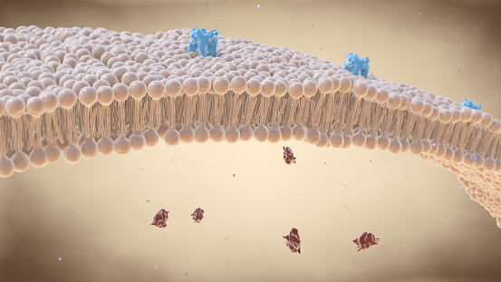
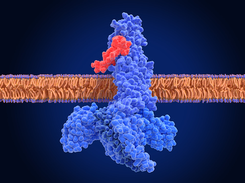
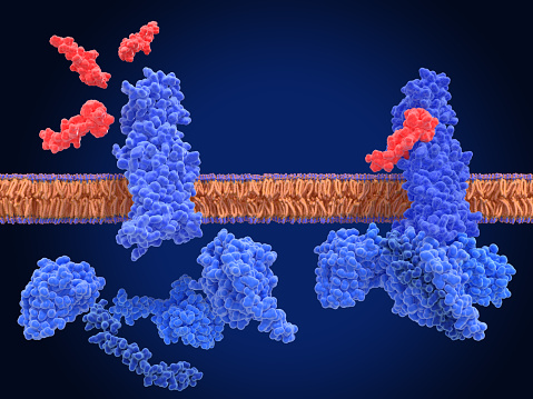
Free Images: "bestof:LDLR pathway.png LDL Receptor Pathway LDL particles blue triangles bind the LDL receptor red on the cell membrane The receptors associate with clathrin green"
pathway-bird-infinite-green-nature-755550.jpg
path-walk-greenery-flowers-red-1342724.jpg
park-pathway-outdoor-road-color-411747.jpg
walkway-hiking-trail-trail-pathway-336624.jpg
autumn-forest-tree-light-sun-path-1075821.jpg
gradient-wallpaper-colors-triangles-1629715.jpg
Sunrise-in-Noosa-national-park-Sunshine-Coast-Queensland-Austr.jpg
sky-blue-outdoor-cloud-photography-1654782.jpg
Trail-through-tall-trees-in-a-lush-forest-Shenandoah-Park.jpg
bridge-brush-trees-path-landscape-843217.jpg
frozen-snow-fall-leaves-forest-1075829.jpg
road-forest-autumn-sky-leaves-1585485.jpg
Ebola_Virus_Particles.jpg
Candy_colored_candles.jpg
Semnalizare engl.svg
Christian_and_Thompson_Figure_5_SVG.svg
EGFR signaling pathway.svg
EGFR signaling pathway (zh-cn).svg
EGFR signaling pathway de.svg
red-eye-green-blue-triangle-41230.svg
red-black-green-blue-white-purple-35240.svg
Ketogenesis.svg
CD8 receptor.svg
gift-loop-bind-decoration-1318358.png
Group d6 id.svg
Fc receptor schematic.svg
Isodynamic from reflections.svg
Intouch Triangle and Gergonne Point.svg
Estrellas.svg
Human genome by functions.svg
Fremantle Prison map.svg
Pathlines streaklines timelines t0-t5.svg
Pathlines streaklines timelines.svg
Insulin glucose metabolism ZP.svg
Insulin glucose metabolism ZP-ca.svg
Flag of the Command of a Cavalry Brigade of the Royal Hungarian Defence Forces (1928).svg
DRAM.svg
Z Music Television - Joe Gow Study.svg
Mathematical-polar-equation-flowers.svg
Zeppelin-LZ-127 internal and gas cells opaque.svg
Zeppelin-LZ-127 internal and gas cells.svg
Positionskarte der Radrouten des Flaeming-Skate in Brandenburg (Transparenter Hintergrund).svg
Triangle-right-abchpq.svg
portugues-sexo_alemão-sex_ingles-sex_africano-seks_russo-секс_albanes-sex_chines-性別_bulgaro-секс_croata-spol_gales-rhyw_coreano-섹스_frances-sexe_japones-セックス_filandes-sukupuolesta_arabe-الجنس_prontos_ai_tens_em_15_linguas_a_p.jpg
LDLR pathway.png
NanoLDLs.jpg
LDLR 1N7D.png
LDLR Binding Module5 1ajj.PNG
PDB 1ijq EBI.jpg
PDB 1n7d EBI.jpg
Monoamine receptor effects.png
1FYT T-cell receptor and HLA class II complex.png
Nodal signaling.jpg
PPARg RXRa 3E00.png
Beta-2 adrenergic receptor.png
PDB 1j8e EBI.jpg
PDB 1f8z EBI.jpg
PDB 1d2l EBI.jpg
PDB 1ldr EBI.jpg
PDB 1k7b EBI.jpg
PDB 1ajj EBI.jpg
PDB 1ldl EBI.jpg
PDB 1f5y EBI.jpg
PDB 1cr8 EBI.jpg
PDB 1d2j EBI.jpg
PDB 1jrf EBI.jpg
PDB 1hj7 EBI.jpg
PDB 1hz8 EBI.jpg
PDB 1xfe EBI.jpg
PDB 1i0u EBI.jpg
PDB 2fcw EBI.jpg
PDB 3bps EBI.png
AhRSignaling colour.png
PDB 1v9u EBI.jpg
EGFR signaling pathway.png
EGF Receptor.jpg
TCR-MHC II.png
CGRP-receptor+RAMP1 3N7P.png
CD8 receptor.png
CA1_hippocampus.jpg
Bzr pm.png
PNN.png
CD4 receptor.png
UH-301-3D-balls.png
UH-301-3D-spacefill.png
Renin ang pathway 2.jpg
Structure of LDLR Family.png
PDB 1rew EBI.jpg
PDB 1es7 EBI.jpg
Gel_Reductor.jpg
RECEPTOR.GIF
PDB 1bm1 EBI.jpg
PDB 2brd EBI.jpg
PDB 1brd EBI.jpg
PDB 1m0k EBI.jpg
PDB 1m0l EBI.jpg
PDB 1m0m EBI.jpg
Terms of Use   Search of the Day