Click Here for More Images from iStock- 15% off with coupon 15FREEIMAGES 
Accurate xray image of human skeletal system with adult male skeleton and body contours on blue background 3D rendering illustration. Anatomy, medical, healthcare, science, osteology, concept.
Medical student standing next to a skeleton
human male skeleton rotating on black background, skeletal system, medically accurate, bone structure Skull, spine, rib cage, pelvis, joints, full body anatomy, 3d render
Image of head-to-toe scan of a fit woman and muscular man body. Comparison of male and female body. MRI scanning of the human body skeletal and muscular systems. You can see the animation movie of this image from my iStock video portfolio. Video number: 1359403986
The true ribs include rib pairs 1-7, with each rib articulating posteriorly to the thoracic vertebrae and anteriorly to the sternum via costal cartilages
3DCG anatomical skeleton
A digital radiograph is used to produce a high-precision analog X-ray film. It is used for diagnostics in the field of internal and external diseases.
A female doctor explains an x-ray.
Accurate xray image of human skeletal system with adult male body contours on black background 3D rendering illustration. Anatomy, osteology, medical, healthcare, science concept.
The human skeleton is the structural framework of the body, providing support, protection for internal organs, and allowing for movement. It is composed of bones, joints, and cartilage.
Xray view of a patient with an injured spine and suffering. / You can see the animation movie of this image from my iStock video portfolio. Video number: 1453180480
Human skeleton, opened mouth, copy space
3D illustration of male human body skeletal system.
3D Illustration Concept of Human Skeleton System Axial Skeleton Bone Joints Anatomy
Male Human skeleton
Male skeleton and transparent skin multiple poses black background  isolated 3d render
Human skeleton for demonstration made of plastic
Accurate human skeletal system 3D rendering illustration. Anterior, lateral, posterior and three-quarter front views of full skeleton with male body contours on blue background. Anatomy, osteology concepts.
Radiography is a method of examination that gives an image of an internal organ using X-rays, while magnetic resonance imaging (MRI), computed tomography (CT) and ultrasound imaging (ultrasound) can also be used.
The intertransverse ligaments comprise sheets of connective tissue that extend from one transverse process to the next 3d illustration
\
3D Rendering Skeletal Anatomy Posterior Anterior
MRI scan review of one of the most difficult positions in gymnastics. The image is showing the skeletal structure of spagat position on the mat. Woman performing a challenging gymnastics position with her legs spread wide on the floor. Woman health and yoga. A human anatomy view created with x-ray images. / You can see the animation movie of this image from my iStock video portfolio. Video number: 1359978210
This 3d animation shows the pectoralis minor muscles on a male human boby
back view of human body 3d
Front and back views of full human skeleton with male body 3D rendering illustration isolated on black. Anatomy or medical diagram with each bone or group of bones labeled with colors.
A human replica skeleton with arms raised on a black background. Focus selective on breast.
3d rendered, medically accurate illustration of a male skeleton system
Human skeleton model of a doctor: chest and head
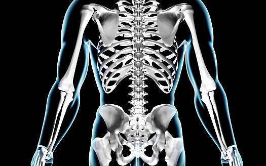
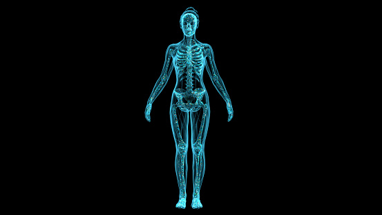
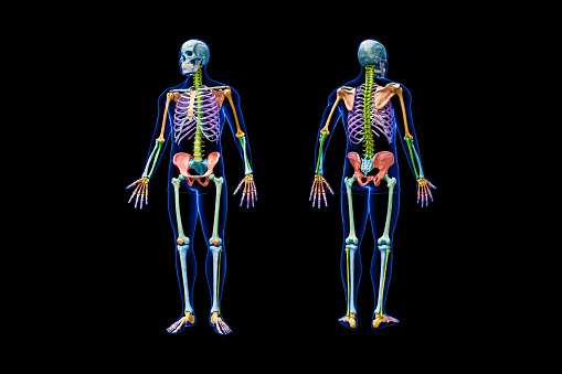
Free Images: "bestof:Human skeleton front hu.svg női csontváz áttekintő ábrája A piros nyilak egy-egy csontot mutatnak a kékek több csontból álló részeket left thumb"
Human skeleton front hu.svg
Frontotemporale.PNG
Terms of Use   Search of the Day