Click Here for More Images from iStock- 15% off with coupon 15FREEIMAGES 
Leiden leaf c.s. under microscope
3d rendering of transparent molecule shape on green color background.
Host cells with spores (mold) are inside wood under the microscope for education.
Bone marrow plasma cells .Plasma cell neoplasms are diseases in which the body makes too many plasma cells. Plasma cell neoplasms can be benign or malignant. Diagnosed with Immunostain CD138 positive.
Squalane is a hydrocarbon derived by hydrogenation of squalene. 3d illustration
Photomicrograph of filamentous blue-green algae, Oscillatoria species. Each band along the stand is one cell. This filament is very short probably having recently parted from a longer one. Filaments are typically many times longer. This algae is mobile, it moves and twists through the water. San Francisco Bay, California, USA. Live specimen. Wet mount, 40X objective, transmitted brightfield illumination. Note - motion blur of live specimen, very shallow depth of field, chromatic aberration and uneven focus are inherent in light microscopy.
Molecular structure formula. Concept. 3D Render
Motor Neuron cells under the microscope
Aerial view of loyster farm by Ardara, County Donegal - Ireland.
3D rendering of microscopic particles and bubbles
The study of plant tissues under the microscope in the laboratory.
\
An antibody, also known as an immunoglobulin, is a large, Y-shaped protein used by the immune system to identify and neutralize foreign objects such as pathogenic bacteria and viruses. 3D cartoon and Gaussian surface models with differently colored chains, white background.
Quantum Gravity 3d rendered image of a comprehensive exploration of Quantum Gravity theories. String theory, loop quantum gravity (LOG), causal dynamical triangulation, twistor theory, gravitational waves, spacetime, gravity waves, quantum mechanics, quantum entanglement concepts.
Cross section leaf of plant under the light microscope view for botany education.
plant Zea Stem C.S. under light mircoscope with white background
Blue colored stem cells of a lentil plant under the microscope.
Candida Auris Cells.  3D Render
Backgrounds of Bacteria and Mold under the microscope in Lab.
Photomicrograph of flatworm,
Thyroid cancer: Microscopic image of Metastatic papillary carcinoma of thyroid. Reactive change. Lymph node carcinoma.
Blastomycosis is a fungal infection caused by inhaling Blastomyces dermatitidis spores. If it involves only the lungs, it is called pulmonary blastomycosis. Only about half of people with the disease have symptoms, which can include fever, cough, night sweats, muscle pains, weight loss, chest pain, and feeling tired.
Algal research, energy and healthcare treatment biotechnology, photobioreactor in medical science laboratory, algae fuel biofuel industry, coronavirus covid-19 vaccine, sustainable eco living
A molecular model of Bisphenol A or BPA.  It is a common building block in the production of many epoxy resins and plastics including polycarbonate plastic. As such it is present in a huge range of food packaging and household products. There are now concerns about its health effects, particularly its effect on children, infants and fetuses.   Isolated on white.
Close up of cristal beads
Dna structure interior body human strong glass clear crystal test tube laboratory connection background transparent reflective metal surface liquid inside. Medical science concept. 3D Illustration.
Cross-section Dicot, Monocot and Root of Plant Stem under the microscope for classroom education.
The three-dimensional structure of proteins is represented in a variety of conformations. This image is based on the ribbon structure of a protein. It is suitable for biology notebooks and book covers.\nThis image has not been created by AI.
Baryte or barite is a mineral consisting of barium sulfate. The baryte group consists of baryte, celestine, anglesite and anhydrite. 3d illustration
Liver Fluke Section under light microscope with white background
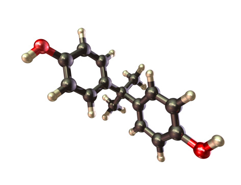
Free Images: "bestof:Hoechst 33258 Structure.gif Structural formula of fluorescent DNA stain Hoechst 33258 Copy since english Wikipédia Image Hoechst 33258 Structure gif by Ygonaar"
Eiffel_Tower_Closeup.jpg
Eiffel closeup.jpg
Lunar libration with phase Oct 2007.gif
Lunar libration with phase2.gif
Color coded racetrack large channel.gif
Sun_Setting_at_Seattle_Airport.jpg
Muybridge_race_horse_animated_184px.gif
Moenitarri_warrior_in_the_costume_of_the_dog_danse_0056v.jpg
Jean-Baptiste-Jules_Bernadotte,_Prince_de_Ponte-Corvo,_roi_de_Suède,_Maréchal_de_France_(1763-1844).jpg
Al-Idrisi's_world_map.JPG
Spectra of Hoechst dyes.svg
Structure of Hoechst dyes.svg
Bisbenzimide.svg
Pyrazophos 200.svg
Earthquake_wave_paths.svg
Average prokaryote cell- en.svg
Aliflurane structure.svg
DNA chemical structure Ar.svg
Taxol structure.svg
DVB-H-framestructure.svg
Sphingolipid de.svg
Busulfan.svg
Methcathinone skeletal.svg
Daptomycin Ball et al.svg
5-amino-2-hydroxybenzoic acid 200.svg
Proteinstructur-ar.svg
Bicuculline.svg
portugues-sexo_alemão-sex_ingles-sex_africano-seks_russo-секс_albanes-sex_chines-性別_bulgaro-секс_croata-spol_gales-rhyw_coreano-섹스_frances-sexe_japones-セックス_filandes-sukupuolesta_arabe-الجنس_prontos_ai_tens_em_15_linguas_a_p.jpg
Binary tree structure.svg
Flag of Dubai.svg
Basal_Ganglia_and_Related_Structures.svg
Hurricane-profile-blank.svg
Iberia Late Bronze.svg
Endomembrane system diagram en (edit).svg
Silica.svg
Animal mitochondrion diagram en.svg
George Curzon2.jpg
Animal mitochondrion diagram en (edit).svg
Animal cell structure en.svg
Hurricane-profile-pt.svg
MaxwellBoltzmann-mk.svg
Hurricane-profile-en.svg
Max_Valier_in_Rocket_Car_(1931).jpg
Tryptophan biosynthesis (en).svg
Design_Analysis_of_the_P-38_Lightning.png
Mesalazine structure.svg
Bombykol Structure V.1.svg
Perfluorobutylamine Structure V.1.svg
Prazosin Structure V.1.svg
Isobutane structure.svg
Tegaserod structure.svg
Dabigatran structure.svg
Ivabradine structure.svg
Laquinimod structure.svg
h2o-water-molecule-580992.png
Semagacestat structure.svg
Diphenyl diselenide Structural Formula V.1.svg
Monothioacetal Structural Formula V.1.svg
Dithioacetal Structural Formula V.1.svg
Hexyl Amine Structural Formula V.1.svg
Ethylmethylthiambutene structure.svg
Losartan structure.svg
Dabigatran etexilate structure.svg
Topaquinone structure.svg
IC-26 structure.svg
MDMA (simple).svg
Ethocybin.svg
DNA intercalation.svg
dna-biology-science-helix-protein-163710.jpg
Cat silhouette.svg
Ethylene.svg
Epigallocatechin.svg
Horseracingicon.svg
MRAM-Cell-Simplified.svg
Diphenylpyraline.svg
MDMA image.svg
Triamcinolonhexacetonide.svg
Trastuzumab emtansine.svg
Plant cell structure svg labels.svg
Harmaline structure.svg
Pethidine structure.svg
3-fluoromethcathinone structure.svg
Prednisone dropshadow.svg
Maltose structure.svg
EcologyTheta.svg
Lawesson's reagent.svg
Methyl isocyanide.svg
Allyl isocyanide.svg
Earth-cutaway-schematic-english.svg
Plant cell structure-en.svg
Paramethoxyethylamphetamine.svg
Levorphanol2.svg
Dihydromorphine.svg
Diprenorphine.svg
Dipropanoylmorphine.svg
Nicocodeine.svg
Nicodicodeine.svg
Nicomorphine.svg
Terms of Use   Search of the Day