Click Here for More Images from iStock- 15% off with coupon 15FREEIMAGES 
Plasmacytoid urothelial carcinoma (PUC) is a unique variant of invasive urothelial carcinoma characterized by tumor cells that exhibit a striking resemblance to plasma cells, and it is known to have an aggressive behavior.
Bladder Cancer: High grade transitional cell carcinoma (TCC), Urothelial carcinoma
Microscopic photo of a thin section of calcareous tufa of Holocene age.
Micrograph of amoeba.  Live specimen. Wet mount, 40X objective, transmitted brightfield illumination. Note - motion blur of live animal, very shallow depth of field, chromatic aberration and uneven focus are inherent in light microscopy.
Thyroid cancer: Microscopic image of Metastatic papillary carcinoma of thyroid. Reactive change. Lymph node carcinoma.
Backgrounds of Bacteria and Mold under the microscope in Lab.
Liver Fluke Section under light microscope with white background
Lung cancer, 3D illustration and photo under microscope. Light micrograph showing lung adenocarcinoma
anaplastic thyroid carcinoma with heterologous differentiation (leiomyosarcoma)
PAPS Smear study of a young women under microscopy showing Atrophic changes in uterus which is unusual at young age.
Natural agate surfaces, suitable for backgrounds and wallpapers.
Stratified flat epithelium under microscope
Photomicrograph of two rotifers. Rapidly rotating cilia at top of heads, internal organs visible. Live specimen. Wet mount, 10X objective, transmitted brightfield illumination.
Kidney cancer, sample collected by CT guided FNA from renal mass. Micrograph of small cell carcinoma, show granular salt-pepper chromatin, background inflammatory cells and blood, selective focus
plant Pine Cone-Female L.S. under light mircoscope with white background
Bacteria Gram positive in sputum gram stain.red background.
Diffuse large B-cell lymphoma, or DLBCL, is a cancer that starts in white blood cells called lymphocytes.
Photomicrograph of Histological analysis of Intramedullary SOL showing Astrocytoma, WHO grade-4
Slice of natural crystal for patterns and textures use with high resolution.
Microscopic image of onion root tip cells undergoing mitosis. Anaphases and metaphases. 1000x magnified
Squash preparation of plant cells stained to reveal chromosomes, various stages of mitosis are visible. Bright field illumination, light microscopy. This image was taken with the X40 objective lens. Depth of focus is extremely low at this magnification. Smaller apertures give greater depth of focus, but image quality deteriorates and resolution is greatly reduced. The aperture in this image was optimised for contrast and resolution. Chromatic aberration is inevitable in light microscopy, but has been kept to a minimum in this image.
Histological Cerebellum human, Sympathetic ganglion human And Spinal cord human under the microscope for education.
Hemangioma biopsy under light microscopy zoom in different regions
Metastatic plasmacytoma, metastatic undifferentiated sarcoma. Microscopically show cellular material of mostly plasmacytiod cells. atypical mitoses are present. plasma cell tumor.
High-grade serous ovarian carcinoma with SET features
Light microscope micrograph showing mitosis in onion cells of the root meristem. At bottom, is a cell in anaphase. On the top right corner, a prophase.
\
Invasive squamous cell carcinoma of larynx, grade II
Captured by a scientific microscope and Canon 5D Mark IV
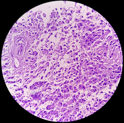
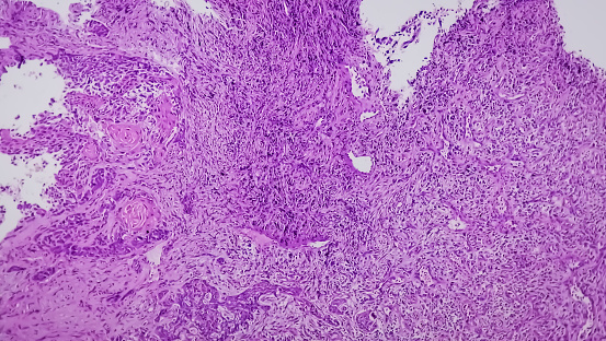
Free Images: "bestof:Hepatocellular carcinoma 1.jpg Hepatocellular carcinoma This specimen is from a 50ish woman who presented to the hospital with abdominal pain and ascites The"
Hepatocellular carcinoma 1.jpg
Hepatocellular carcinoma 2.jpg
Adenocarcinoma of the stomach.jpg
Cervical Carcinoma with Adnexa.jpg
Hamartoma of the spleen.jpg
Serous Cystadenocarcinoma of the Ovary.jpg
ChronicBackache1041.png
Terms of Use   Search of the Day