Click Here for More Images from iStock- 15% off with coupon 15FREEIMAGES 
A ball and stick model of Human Insulin. Isolated on black.
Proteasomes are molecular machines for breaking down proteins called proteolysis, only target proteins that have been marked for destruction called ubiquitin to the target protein, 3d rendering
3d rendered Gastric inhibitory polypeptide (GIP, glucose-dependent insulinotropic peptide) endocrine protein hormone, 3D illustration
The three-dimensional structure of proteins is represented in a variety of conformations. This image is based on the ribbon structure of a protein. It is suitable for biology notebooks and book covers.\nThis image has not been created by AI.
p53 (blue) tagged with ubiquitin (yellow) is degraded into small peptides by a proteasome (violett). p53 prevents cancer formation and acts as a guardian of the genome. Mutations in the p53 gene contribute to about half of the cases of human cancer.
3d rendering of transparent molecule shape on green color background.
An antibody, also known as an immunoglobulin, is a large, Y-shaped protein used by the immune system to identify and neutralize foreign objects such as pathogenic bacteria and viruses. 3D cartoon and Gaussian surface models with differently colored chains, white background.
ATP Synthase Protein - 3d rendered image. Medical microbiology concept.  Structure and conformational states of the bovine mitochondrial ATP (Adenosine triphosphate) synthase. Chemical energy is synthesized in eukaryotic cells primarily. Hydrolase. Hologram view. HUD, FUI
Human insulin hormone molecule, space filled model, 3D illustration. Drug in diabetes treament
3d Rendering Molecular Structure,Ozone,Hydrogen and oxygen
Inside protein molecule
Science background with molecule or atom
Nirmatrelvir inhibits the 3C-like protease of the SARS-CoV 2 virus. The coronavirus 3CL hydrolase enzyme, also known as the main protease, is essential for the maturation of the virus.
Molecular structure formula. Concept. 3D Render
A ball-and-stick molecular model of the hormone Orexin, also known as Hypocretin.  Orexin is involved in the regulation of the sleep/wake cycle and it stimulates apetite, wakefulness and energy expenditure. It has been linked with Alzheimer's disease.  Isolated on white.
3D cartoon and Gaussian surface models, PDB 6vg8, white background
Calcidiol molecule, also called calcifediol, major circulating metabolite of vitamin D3 produced in liver, 3D illustration
Endosomes - 3d rendered image of Endosomes are membrane-bound organelles. They are parts of endocytic membrane transport pathway.  Endomembrane system in cells. Bilayer Lipid Membrane. Abstract medical illustration. Microscopic view. Microbiologic research concept.
Hyaluronan, hyaluronic acid, HA, hyaluronate molecule, short fragment. Molecular model. 3D rendering. Illustration
Slice of natural crystal for patterns and textures use with high resolution.
From left to right: the NMDA and  AMPA receptors transport calcium cations into neurons after being activated by the neurotransmitter glutamate and  the GABA receptor (right) transport chloride anions after the activation by gamma aminobutyrate. PDB entries: 6wht,  3kg2, 6d6u
DNA A-T Pair
Biological protein and molecule, 3d rendering. 3D illustration.
molecular symbol painted on the wall
CD47 also known as integrin associated protein is a transmembrane protein that in humans is encoded by the CD47 gene. CD47 belongs to the immunoglobulin superfamily and partners with membrane integrins and also binds the ligands thrombospondin-1 and signal-regulatory protein alpha. 3D cartoon and Gaussian surface models, chain id color scheme, based on PDB 2jjt, white background
Endosomes - 3d rendered image of Endosomes are membrane-bound organelles. They are parts of endocytic membrane transport pathway.  Endomembrane system in cells. Bilayer Lipid Membrane. Abstract medical illustration. Microscopic view. Microbiologic research concept.
Multicolored antibodies or immunoglobulin protein structures - 3d illustration
Myxoid liposarcoma.  Myxoid/round cell liposarcoma, or MRCLS, is one of several types of liposarcoma. Liposarcoma is a rare cancer that grows in the cells that store fat in the body. MRCLS usually grows in the arms and legs. These tumors grow slowly, and they can spread to other parts of the body
The NEK-7 independent NLRP3 inflammasome in it's hexameric conformation with ADP (red) bound. Inflammasomes are a promising target for the treatment of autoimmune diseases. PDB entry: 7zgu.
3D realistically coloured hexagonal shapes of connected spheres, background with copy space. Futuristic illustration design template for scientific presentation. Molecular business, science, teamwork concept.
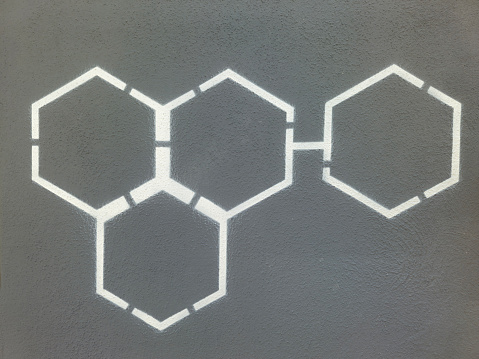
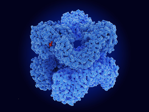
Free Images: "bestof:Helice alpha spire 0.png Hydrogen liaison in alpha protein structure - 2006-03-30 pixeltoo Protein structures"
Fine structure of Ly-alpha.svg
Alpha sheet bonding schematic-color.svg
Hydrogen fine structure.svg
Helice alpha spire 0.png
Helice alpha spire.png
Carboxys shell.png
PDB 1i6a EBI.jpg
PDB 1i69 EBI.jpg
Clostridium perfringens Alpha Toxin.rsh.png
TNFa Crystal Structure.rsh.png
PDB 1snu EBI.jpg
Tonge redo alpha mannosidase.png
Clostridium perfringens Alpha Toxin Rotate.rsh.gif
Calmodulin 1CLL.png
PDB 1wjk EBI.jpg
Proteina-color1.PNG
Aluminium-hydride-H-coordination-3D-balls.png
NH2-Domäne von Kollagen Typ IX, alpha 2.png
RCSB-PDB-1I1S.png
Crystal Structure of the Ku Heterodimer - 15.png
Crystal Structure of the Ku Heterodimer - 16.png
Crystal Structure of the Ku Heterodimer - 17.png
Crystal Structure of the Ku Heterodimer - 2.png
Crystal Structure of the Ku Heterodimer - 20.png
Crystal Structure of the Ku Heterodimer - 22.png
Crystal Structure of the Ku Heterodimer - 23.png
Crystal Structure of the Ku Heterodimer - 26.png
Crystal Structure of the Ku80 - 11.png
Crystal Structure of the Ku Heterodimer - 30.png
Crystal Structure of the Ku Heterodimer - 31.png
Crystal Structure of the Ku Heterodimer - 33.png
Crystal Structure of the Ku Heterodimer - 36.png
Crystal Structure of the Ku80 - 37.png
Crystal Structure of the Ku80 - 38.png
Crystal Structure of the Ku80 - 39.png
Crystal Structure of the Ku80 - 40.png
Crystal Structure of the Ku70 - 41.png
Crystal Structure of the Ku70 - 42.png
Crystal Structure of the Ku70 - 43.png
Crystal Structure of the Ku Heterodimer - 35.png
Bt toxin.jpg
Crystal structure of Equ c 1 dimer.jpg
I-CreI dimer 1.png
HLA-Cw4.png
Mup1 PDB 1i04.jpg
Amino-CORN.png
MupwithSBT.jpg
PPARg RXRa 3E00.png
I-CreI dimer DNA.png
I-CreI dimer DNA 2.png
Terms of Use   Search of the Day