Click Here for More Images from iStock- 15% off with coupon 15FREEIMAGES 
Primary clear cell carcinoma of the pancreas is rare and it is classified as a \
Showing Light micrograph Type of Tissue Human under the microscope in Lab.
PAPS Smear study of a young women under microscopy showing Atrophic changes in uterus which is unusual at young age.
Abstract blue cell pattern, spots, cool, virus, background effect, texture
Human brain anatomy, basal ganglia. 3D illustration showing caudate nucleus (green), putamen (yellow), and lateral ventricles (blue)
A microscope slide showing a stained section of a human bowel, or small intestine.  It was taken through microscope lens that has a central focus through a thin layer of glass.
Quantum Gravity 3d rendered image of a comprehensive exploration of Quantum Gravity theories. String theory, loop quantum gravity (LOG), causal dynamical triangulation, twistor theory, gravitational waves, spacetime, gravity waves, quantum mechanics, quantum entanglement concepts.
Sound speaker and structure of the human ear. Influence of loud sound on hearing.
Kidney cancer: Clear cell carcinoma. fuhrman nuclear grade-II, Renal growth shows a malignant tumor, renal cell carcinoma in between renal tubules.
WHO grade I menWorld Health Organization (WHO) grade I meningiomas are intracranial extracerebral tumors, in which microsurgery as a stand-alone therapy provides high rates of disease control and low recurrence rates. ingioma.
Tissue of Small intestine (Duodenum), Large intestine Human and Stomach Human under the microscope in Lab.
Wilms tumor, or nephroblastoma, light micrograph, photo under microscope
Close-up shooting of a green agate stone mineral surface isolated on white background. Its a macro shot shows nature beautiful details with back lit. Agate is a kind of natural gemstone. Abstract pattern. Good for Wallpaper texture background. Purple
Human lung pathology under light microscope, The lungs is organs of the respiratory system in human. Human pathology education. Haematoxylin and eosin staining technique slide.
abstract imagery of a fusion of colors and surfaces
Prostate Cancer: Microscopic image of prostatic tissue, adenocarcinoma, malignant neoplasm, atypical epithelial cells.
Serous carcinoma is the most common type of ovarian cancer, accounting for approximately 75% of epithelial ovarian cancers.
Hyaline cartilage, Elastic cartilage and Bone Human under the microscope in Lab.
Acute ordinary hepatitis liver biopsy under light microscopy
Eye disease, eye cancer, 3d illustration
Model - Object, Listening, Blackboard, Body Part, Human Body Part
The prostate is both an accessory gland of the male reproductive system
Magnetic resonance images of the brain (MRI brain). Computed resonance imaging of the human brain.
Human neuron cells with brain. 3d illustration
Cardiac amyloidosis is a condition where faulty proteins build up in your heart. You can inherit this condition, or it can develop on its own (usually later in life). As faulty proteins accumulate, your heart struggles to pump, ultimately leading to heart failure and death.
Histological Pancreas human, Liver human, Vermiform appendix human and Kidney Human under the microscope for education.
Kidney proximal tubule epithelial cloudy SWELLING under different area in light microscopyKidney proximal tubule epithelial cloudy SWELLING under different area in light microscopyKidney proximal tubule epithelial cloudy SWELLING under different area in light microscopy
Different germs in the human intestines called microbiota - 3d illustration
Senior Caucasian man wearing hearing aid
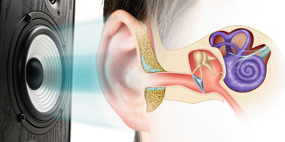
Free Images: "bestof:Hearing mechanics cropped - Acoustic radiation.jpg Medial geniculate nucleus to Primary auditory cortex 内側� 状体から� 次聴覚皮質へ� � http //www"
Terms of Use   Search of the Day