Click Here for More Images from iStock- 15% off with coupon 15FREEIMAGES 
Leiden leaf c.s. under microscope
Rare Scorpion (Arachnida) inclusion in Burmese amber, Myanmar, Cenomian, Upper Cretaceous, approximately 100 million years ago, age of dinosaurs. Image taken with extreme macro and focus stacking technique.
Epidermis. Electron microscope micrograph showing a keratinocyte of spinous layer. The epithelial cell has a polygonal shape, central nucleus with nucleolus, cytoplasm full of keratin filament bundles, and numerous dark desmosomes crossing the intercellular spaces.
Bone marrow plasma cells .Plasma cell neoplasms are diseases in which the body makes too many plasma cells. Plasma cell neoplasms can be benign or malignant. Diagnosed with Immunostain CD138 positive.
Microalgae under microscopic view, green algae, cyanobacteria, phytoplankton, diatom, algae mix collage background
Photomicrograph of filamentous blue-green algae, Oscillatoria species. Each band along the stand is one cell. This filament is very short probably having recently parted from a longer one. Filaments are typically many times longer. This algae is mobile, it moves and twists through the water. San Francisco Bay, California, USA. Live specimen. Wet mount, 40X objective, transmitted brightfield illumination. Note - motion blur of live specimen, very shallow depth of field, chromatic aberration and uneven focus are inherent in light microscopy.
amazing inhabitants of the microworld under a microscope
plant Zea Stem C.S. under light mircoscope with white background
Penicillium mold fungi, 3D illustration and photo of colonies grown on nutrient medium
Rat tailed maggot photographed in a studio
Planaria worm in pond water
Trematode parasite under a microscope
Chaetoceros is the largest genus of marine planktonic diatoms, phytoplankton
Red skunk cleaner shrimp - Lysmata Amboinensis
Planarian parasite (flatworm) under microscope view.
Monotomidae, Coleoptera Fossil in Burmese amber of Cenomanian era, 100 million years ago, from the state of Myanmar, extreme macro shot
Photomicrograph of algae, Cosmarium turpinii, a desmid. Live specimen. Wet mount, 40X objective, transmitted brightfield illumination.
Freshwater aquatic zooplankton and algae under microscope view
Autumn maple leaves background. Colorful maple leaves background. Autumn maple leaves background
A Banded Demoiselle poised on dead grass head in sunlight
Artemia Salina - Brine Shrimp are food of Seahorses and many other Saltwater Animals
mosquito larva egg under light microscopy
Egg  clump of the rare and endangered Kloof frog, also known as the Natal diving frog, or Boneberg's frog (Natalobatrachus bonebergi) hanging on vegetation above a slow moving stream in a forest
Astragalus bolanderi is a species of milkvetch called Bolander's milkvetch. McGee Creek Canyon, Mono County,  Sierra Nevada Mountains, California.
The larvae of moths are covered with bristles and look terrible
Pleasant Chelidonura Chelidonura amoena occurs in the tropical Indo-West Pacific from Japan down through the Philippines, Malaysia, Indonesia, both east and west coasts of Australia to Hawaii ina depth range from 2-22m, max. length 5.5cm. The carnivore species is often found crawling on coral during daylight hours, feeding on small flatworms which are sometimes quite common in reef environments. This specimen was found on a Encrusting Sponge Clathria sp. on a ship wreck, a post WWII wreck, the Hafa Adai Ferry in Palau 7°19'12.4 N 134°26'39.78 E at 13m depth.
Pine mature wood cross section. Light microscope slide with microsection of an evergreen conifer in the genus Pinus. Plant anatomy. Biology. Photo.
Brown butterfly lying on the ground
Microscopic of paramecium and amoeba
Tiny dragonfly (Zygoptera) perched on a leaf
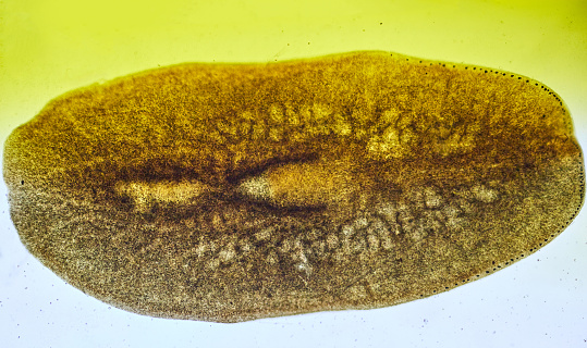
Free Images: "bestof:Gyrocotyle rugosa.png en Internal morphology of Gyrocotyle rugosa Plathelminthes Gyrocotyloidea Spencer W B 1889 The anatomy of Amphiptyches urna Grube and"
Gyrocotyle_rugosa.png
Bolma rugosa.JPG
Everything_for_the_garden_(15768573694).jpg
Terms of Use   Search of the Day