Click Here for More Images from iStock- 15% off with coupon 15FREEIMAGES 
Section of epididymis under light microscope with white background
Enlarged Prostate organ gland as Benign Prostatic Hyperplasia as a urology medical illustration concept for part of the male reproductive anatomy and lower urinary tract symptoms.
Computational tomography image of the abdominal and pelvic regions.
Tissue of Small intestine (Duodenum), Large intestine Human and Stomach Human under the microscope in Lab.
Human Eye anatomy photo taking with Retina Camera.
Magnetic resonance images of the brain (MRI brain). Computed resonance imaging of the human brain.
Anatomy of human eye. medical concept. 3d illustration
plant Flower bud of Lilium brownii var.viridulum T.S.\nunder light mircoscope with white background
3d illustration of a cross section of the human eye with explanations and inscription
MRI scan of veins around the heart
The inner layer of the eye, or retina, is similar to film in a camera. It receives light from an image we are looking at, and converts that light into electrical impulses which are sent through the fibers of the optic nerve to the brain.
Adenocarcinoma, predominantly papillary type, lung
This 3d illustration shows a section of the human eye
skin science 3d
Selective Focus of CT Chest AXIAL MIP view for diagnostic Pulmonary embolism (PE) , lung cancer and covid-19.
Radiology report. On the right, lateral breast there are two intramammary lymph nodes with fatty hilum measuring 0.5 and 0.6 cm respectively. No suspicious breast mass or axillary lymphadenopathy. Moderately dense breasts with 50 to 75 % glandular tissue.  Bi-RADS 2 : benign findings.
Artificial vision concept. Chip with human eye anatomy. 3d illustration
Ct image of human lung on computer monitor in radiology clinic. A thorax ct image of patient with covid-19 disease is seen. Shot indoor with a full frame mirrorless camera.
brainstem is the bottom, stalklike portion of your brain.
High-grade serous ovarian carcinoma with SET features
Showing Light micrograph Type of Tissue Human under the microscope in Lab.
MRI of the prostate gland, revealing an enlarged size, aids in diagnosing tumors, guiding treatment decisions, and monitoring prostate health.
CT angiography of lower limb..artery occlusive disease
A scanned page of an anatomy textbook from the 19th century.
The image depicts a near median section through the nose and nasal passages. Details include nasal cavity, soft and hard palate, uvula, eustachian tube and pharyngeal tonsil. White background.
Human eye anatomy. Inner structure. 3d illustration
WHO grade I menWorld Health Organization (WHO) grade I meningiomas are intracranial extracerebral tumors, in which microsurgery as a stand-alone therapy provides high rates of disease control and low recurrence rates. ingioma.
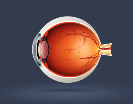
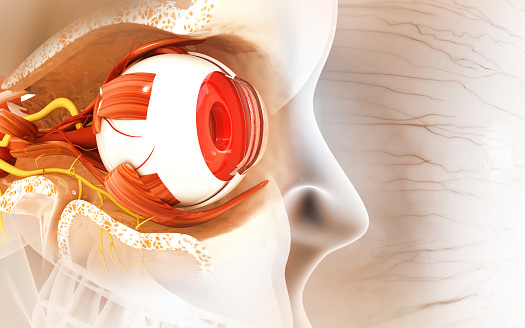
Free Images: "bestof:Gray877.png en Gray's Anatomy plate Diagram of the blood vessels of the eye as seen in a horizontal section Leber after Stöhr <br> Course of vasa centralia"
Gray1158.png
Gray399_es.svg
Gray5_de.svg
Gray5_nl.svg
Gray728.svg
Gray399.svg
Arterial System en.svg
Circulatory System fr.svg
Gray834.svg
Gray5.svg
Temporo-parietal_junction_ja.svg
Venous system en.svg
Arterial System fr.svg
Gray5-ja.svg
Gray770-pt.svg
Circulatory System en.svg
Gray834_es.svg
Gray5_-_es.svg
Gray769-pt.svg
Circulatory System no tags.svg
Gray814_pl.svg
Schematic diagram of the human eye multilingual.svg
Schematic diagram of the human eye en.svg
En-elbow joint.svg
Gray1130.svg
Gray72-en.svg
Gray72-de.svg
Gray770-en.svg
Gray727.svg
Gray1193.svg
Gray1167.svg
Gray477.svg
Gray38.svg
Gray726.svg
Gray1199_pl.svg
Gray37.svg
Gray63.svg
Gray1199_en.svg
Стомах.svg
Gray1157.png
Venous system fr.svg
Culex restuans larva diagram num.svg
Gray1046.svg
Brain_diagram_ja.svg
Gray1167_de.svg
Gray726-Brodman.svg
Gray726-Brodman-prefrontal.svg
Gray1046_pl.svg
Gray506_pl.svg
Gray1193_pl.svg
Gray1167_pl.svg
Brain_diagram_without_text.svg
Gray511.svg
Gray506_es.svg
Gray1167_fi.svg
Gray769-en.svg
Gray1167 es.svg
Gray1095_-_pl.svg
Gray770-es.svg
Gray506-ar-p.svg
Gray799.svg
Lobes_of_the_brain_el.svg
Gray727_el.svg
Gray1236_pl.svg
Gray1235_pl.svg
Gray1199_labeled.svg
Gray511-es.svg
Gray812and814_es.svg
Gray769-es.svg
Gray67.svg
Gray512.svg
Diagram of influenza infection symptoms EN.svg
Diagram of swine influenza symptoms EN.svg
Gray1095.svg
PD Diagram of swine flu symptoms EN.svg
Receptive field.svg
Placenta.svg
Placenta_ja.svg
Placenta_ko.svg
Placenta_de.svg
Placenta_ar.svg
Gray1154.png
Gray588.png
Gray1155.png
Gray1155-IT.png
Gray506_de.svg
Gray1220.svg
Gray424.svg
Gray924.svg
Gray413_blank.svg
Gray1235_(English).svg
Gray828_es.svg
Gray506_cs.svg
Gray646.svg
Gray722_refined.svg
Gray440_es.svg
Gray580.svg
Cross section jellyfish en.svg
Gray1220-ru.svg
Gray506.svg
Terms of Use   Search of the Day