Click Here for More Images from iStock- 15% off with coupon 15FREEIMAGES 
Spike glycoprotein homotrimer, a main target for neutralization antibody, to bind its receptor, ACE2, and mediate membrane fusion and virus entry. 3D cartoon and Gaussian surface models, white background.
The three-dimensional structure of proteins is represented in a variety of conformations. This image is based on the ribbon structure of a protein. It is suitable for biology notebooks and book covers.\nThis image has not been created by AI.
orange: RNA primer; red: RNA template; light blue: proofreading riboexonuclease; mid blue: helicase; dark blue: RNA directed RNA polymerase; violet: non structural proteins (NSPs).
Molecular model of amylose, one of the two sugar components in starch.
3d rendering of ubiquitin is attached to target proteins by a process called ubiquitination
A ball and stick model of a molecule of gamma-Cyclodextrin. Composed of eight glucose sugar molecules bound into a ring structure, cyclodextrins are easily made from simple starch. The large ring can bind smaller molecules within it, making Cylodextrins useful in drug-delivery applications and also to clean up toxins (such as heavy metals) in the environment. Currently, gamma-Cyclodextrin is being considered as a structure which can bind to and hold carbon dioxide gas for carbon capture and storage as a way to manage the effect of excess carbon dioxide on the climate.
Copper(II) sulfate with molecular structure. Chemical ingredient used in medical and public health issues.
Top view of various kinds of vegan protein sources like tofu, tempeh, soy beans, soy milk, mushrooms, wakame, lentils, peanuts, spinach and chick peas. All the objects are at the borders of the image and at the center is written \
Collagen protein molecule. Molecular model. 3D rendering. Illustration
Covid-19 Coronavirus Variants Omicron Delta Gamma Beta Alpha Pandemic Virus Disease Mutation
From left to right: the NMDA and  AMPA receptors transport calcium cations into neurons after being activated by the neurotransmitter glutamate and  the GABA receptor (right) transport chloride anions after the activation by gamma aminobutyrate. PDB entries: 6wht,  3kg2, 6d6u
3D realistically coloured hexagonal shapes of connected spheres, background with copy space. Futuristic illustration design template for scientific presentation. Molecular business, science, teamwork concept.
Structure of novel coronavirus spike receptor-binding domain (pink) complexed with its receptor ACE2 (blue), surface modeel, white background
Chemical formula of Progesterone
Multicolored antibodies or immunoglobulin protein structures - 3d illustration
29 October Republic Day of Türkiye text on a red shiny sphere with a partial Turkish flag on white background. Easy to crop for all your print sizes and social media needs.
Molecular model of insulin hormone, 3D illustration. It is produced by pancreas and takes part in glucose and lipid metabolism
Infinity sign made out of multiple spheres and circular shapes against grey background.
Bacterial multidrug efflux pumps (E-CmeB) expel antibiotics  from inside the bacterium (bottom): ciprofloxacin (yellow), chloramphenicol (violet),  ampicillin (red).
Inside protein molecule
Hyaluronan, hyaluronic acid, HA, hyaluronate molecule, short fragment. Molecular model. 3D rendering. Illustration
A ball and stick model of Eicosapentaenoic Acid or EPA. It is an omega-3 polyunsaturated fatty acid. It is introduced into the diet by eating oily fish. Isolated on white.
c-Myc and Max transcription factors bound to DNA. 3D illustration. Atoms shown as spheres with conventional color coding.
molecular structure digital concept
Microcrystalline wax with chemical formula on white laboratory table.
Chemical formula of Testosterone
3D cartoon and Gaussian surface models, PDB 4bci, white background
Molecular model of amylose, one of the two sugar components in starch
Monosodium glutamate, MSG or sodium glutamate molecules. MSG is used in cooking as a flavor enhancer with an umami taste 3d rendering
A ball and stick model of Human Insulin. Isolated on black.
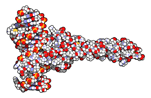
Free Images: "bestof:Glicoprotein.svg N-linked protein glycosylation N-glycosylation of N-glycans at Asn residues Asn-x-Ser/Thr motifs in glycoproteins en Image Glicoprotein jpg"
IC10_BVHa.jpg
Glicoprotein.svg
Lipid raft organisation scheme.svg
2node chain.svg
Node chain.svg
X dominant affected mother.svg
X dominant affected father.svg
Haystacks1989.jpg
Riassorbimento bicarbonati e respirazione cellulare.svg
Style and stigma shape in color-01 all types from APWeb glossary.svg
Haystacks_at_Chailly_at_Sunrise,_1865,_oil_on_canvas,_11_7-8_x_23_3-4_inches_(30_x_60_cm),_San_Diego_Museum_of_Art.jpg
Style and stigma shape in color-03 truncate (truncado).svg
Style and stigma shape in color-08 lobed 1 (estigma lobado 1).svg
Style and stigma shape in color-09 lobed 2 (estigma lobado 2).svg
Style and stigma shape in color-02 not differenciated (indiferenciado).svg
Style and stigma shape in color-16 stylar beak 1 (beak estilar 1).svg
Style and stigma shape in color-10 discoid (discoide).svg
Style and stigma shape in color-11 hairy (con pelos, tricomas).svg
Style and stigma shape in color-12 feathery (plumoso).svg
Style and stigma shape in color-04 filiform (estilo o estigma filiforme).svg
Main protein structure levels bg.svg
Style and stigma shape in color-07 conical (estigma cónico).svg
Style and stigma shape in color-17 stylar beak 2 (con beak estilar 2).svg
Style and stigma shape in color-13 comb-like (como un peine).svg
Style and stigma shape in color-14 fan-shaped (en abanico, flabelada).svg
Wheatstacks_(End_of_Summer),_1890-91_(190_Kb);_Oil_on_canvas,_60_x_100_cm_(23_5-8_x_39_3-8_in),_The_Art_Institute_of_Chicago.jpg
Main protein structure levels zh.svg
Main protein structure levels pl.svg
Style and stigma shape in color-15 petal-like, petaloid (estilo petaloideo).svg
Claude_Monet,_Grainstacks_in_the_Sunlight,_Morning_Effect,_1890,_oil_on_canvas_65_x_100_cm.jpg
Claude_Monet,_Haystacks_on_a_Foggy_Morning,_1891,_Oil_on_canvas.jpg
1290_Grainstack_in_the_Sunlight,_1891,_Oil_on_Canvas,_Private_Collection.jpg
Claude_Monet._Haystack,_Snow_Effects,_Morning._1890._Oil_on_canvas._Museum_of_Fine_Arts,_Boston,_MA,_USA..JPG
Style and stigma shape in color-06 club-shaped (clavado, con forma de garrote).svg
Claude_Monet._Haystack._End_of_the_Summer._Morning._1891._Oil_on_canvas._Louvre,_Paris,_France.jpg
Poss_1288_Grainstack_in_Sunshine,_1891,_Meule_au_soleil,_Oil_on_Canvas,_60_x_100_cm,_Zurich,_Kunsthaus_Zurich.jpg
1278_Wheatstacks_(Sunset,_Snow_Effect),_1890-91,_65.3_x_100.4_cm,_25_11-16_x_39_1-2_in.,_The_Art_Institute_of_Chicago.jpg
Claude_Monet,_Haystack,_Morning_Snow_Effect_(Meule,_Effet_de_Neige,_le_Matin),_1891,_oil_on_canvas,_65_x_92_cm,_Museum_of_Fine_Arts,_Boston.jpg
Vulva symbols.svg
Richard Jordan Gatling - Brady-Handy.jpg
Espinosa,_1645,_A_bouquet_of_flowers_in_a_conch_shell,_with_nuts_and_figs,_surrounded_by_a_wreath_of_flowers_and_fruit.jpg
Full_length_picture,_Kevin_O'Higgins,_Minister_of_Home_Affairs.jpg
Charles Stewart Parnell - Brady-Handy.jpg
T Cell Surface Glycoprotein CD4 - PDB 1CDH.jpg
Deinonychus antirrhopus.jpg
Genetic Code Structure.JPG
Bloodgroup-Blutgruppen-Glyk.jpg
APE1bendingDNA.jpg
PBB Protein PRTN3 image.jpg
PBB Protein HIF1A image.jpg
PBB Protein EGF image.jpg
PBB Protein DUSP22 image.jpg
DEADboxprotein405.jpg
PBB Protein BRCA2 image.jpg
PBB Protein POLA1 image.jpg
PBB Protein MAP3K7 image.jpg
PBB Protein TOMM20 image.jpg
PBB Protein WEE1 image.jpg
PBB Protein TPH1 image.jpg
PBB Protein CDK5 image.jpg
PBB Protein SRC image.jpg
PBB Protein PYY image.jpg
PBB Protein MRPL11 image.jpg
PBB Protein KCNH2 image.jpg
PBB Protein VWF image.jpg
PBB Protein APOA1 image.jpg
PBB Protein NTRK1 image.jpg
PBB Protein NF1 image.jpg
PBB Protein CDH1 image.jpg
PBB Protein SOX6 image.jpg
PBB Protein FOS image.jpg
PBB Protein APP image.jpg
PBB Protein RAN image.jpg
PBB Protein DMD image.jpg
PBB Protein VIM image.jpg
PBB Protein NF2 image.jpg
PBB Protein GCG image.jpg
PBB Protein BCR image.jpg
PBB Protein ATP7B image.jpg
PBB Protein MYH7 image.jpg
PBB Protein POT1 image.jpg
PBB Protein TERF1 image.jpg
PBB Protein TERF2 image.jpg
PBB Protein PML image.jpg
PBB Protein MTHFD1 image.jpg
PBB Protein TERF2IP image.jpg
PBB Protein ACTA1 image.jpg
PBB Protein SRF image.jpg
PBB Protein CREB1 image.jpg
PBB Protein TMPO image.jpg
PBB Protein WAS image.jpg
PBB Protein PCK1 image.jpg
PBB Protein COL8A1 image.jpg
PBB Protein ARID1A image.jpg
PBB Protein ARID1B image.jpg
PBB Protein AIRE image.jpg
PBB Protein ATF2 image.jpg
PBB Protein SOX2 image.jpg
PBB Protein CEBPA image.jpg
PBB Protein NR1H4 image.jpg
Terms of Use   Search of the Day