Click Here for More Images from iStock- 15% off with coupon 15FREEIMAGES 
Human digestive system and gastrointestinal tract with microvilli of the small intestine or bowel 3D rendering illustration. Anatomy, medical, biology, microbiology, science, healthcare concepts.
Gastrointestinal stromal tumors (GISTs) are the most common mesenchymal neoplasms of the gastrointestinal tract. GISTs arise in the smooth muscle pacemaker interstitial cell of Cajal, or similar cells. Most (66%) occur in the stomach and gastric GISTs have a lower malignant potential than tumors found elsewhere in the GI tract. Micrograph of gastrointestinal stromal tumor of the stomach.
Woman touching stomach painful suffering from stomachache causes of menstruation period, gastric ulcer, appendicitis or gastrointestinal system disease. Healthcare and health insurance concept.
Sagittal section of human stomach
woman's stomach with redness. stomach problems concept
\
Histological Pancreas human, Liver human, Vermiform appendix human and Kidney Human under the microscope for education.
Intestinal microbiome,Probiotics health benefits,Gut bacteria,lactococcus, propionibacterium, bifidobacterium.
Young woman holding her stomach
Squamous cell carcinoma or squamous cell cancer (SCC or SqCC) is a cancer of a kind of epithelial cell, the squamous cell. These cells are the main part of the epidermis of the skin, and this cancer is one of the major forms of skin cancer. However, squamous cells also occur in the lining of the digestive tract, lungs, and other areas of the body, and SCC occurs as a form of cancer in diverse tissues, including the lips, mouth, esophagus, urinary bladder, prostate, lung, vagina, and cervix, among others. Despite sharing the name squamous cell carcinoma, the SCCs of different body sites can show tremendous differences in their presenting symptoms, natural history, prognosis, and response to treatment. Micrograph of squamous cell carcinoma of the head and neck
Human bladder cancer as a urinary anatomical organ disease and malignant cells concept as a 3D illustration cutaway of body anatomy.
Human stomach internal organ design element illustration concept.
Medical x-ray  illustration of stomach cancer - stomach tumor
Unwell mid adult man having stomach pain at home.
Low magnification micrograph of the gastric mucosa showing the surface mucous epithelium, the gastric pits and the fundic glands with many pink parietal cells.
Close-up of a purple Tie Dye pattern on a textile. Can be used as a background. Tie Dye pattern was created by the photographer.
stomach ache. Asian women have abdominal pain, indigestion, gastritis, menstrual cramps, flatulence, diarrhea, distention, colon cancer, belly inflammation problem, suffer food poisoning, abdomen
Serous carcinoma is the most common type of ovarian cancer, accounting for approximately 75% of epithelial ovarian cancers.
Human digestive system anatomy on blue color background. 3d illustration
\
Human Stomach with endoscope and close-up view of bacterium Helicobacter pylori which causes ulcers, stomach ulcer or gastric ulcer, 3d illustration
Doctor holding human Colon anatomy model. Colonic disease, Large Intestine, Colorectal cancer, Ulcerative colitis, Diverticulitis, Irritable bowel syndrome, Digestive system and Health concept
Large Intestine, Problem, Intestine, Stomachache, Irritable Bowel Syndrome
Senior man with stomach pain
Metastatic neuroendocrine tumor. Site: Liver.  Neuroendocrine tumors are cancers that begin in specialized cells called neuroendocrine cells. Neuroendocrine cells have traits similar to those of nerve cells and hormone-producing cells. Neuroendocrine tumors are rare and can occur anywhere in the body.
Parasitic worms in human large intestine, 3D illustration. Enterobius vermicularis and other round worms
Gastric mucosa intestinal metaplasia under microscopy
Human stomach anatomy. 3d illustration
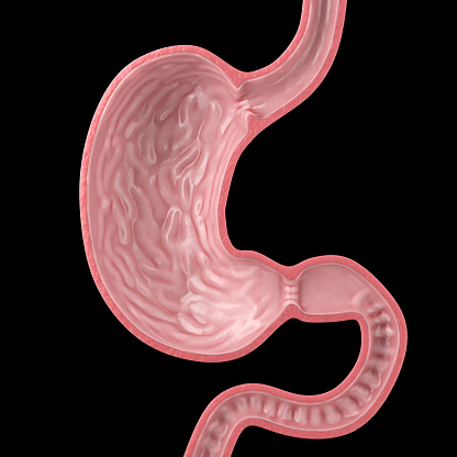
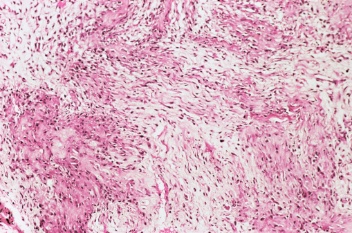
Free Images: "bestof:Gastrointestinal Stromal Tumor (GIST) of Stomach.jpg Gastrointestinal Stromal Tumor of Stomach This 6 5-cm tumor was removed from the stomach of a 68-year-old"
Upper gastrointestinal tract.svg
digestive-organs-stomach-colon-40607.svg
Stomach colon rectum diagram-ca.svg
Stomach colon rectum diagram-bn.svg
Stomach colon rectum diagram-ar.svg
Stomach colon rectum diagram-fr.svg
Stomach colon rectum diagram-is.svg
Stomach colon rectum diagram-th.svg
Stomach colon rectum diagram-ms.svg
Stomach_colon_rectum_diagram_unlabeled.svg
Stomach_colon_rectum_diagram_arrows.svg
Stomach_colon_rectum_diagram-az.svg
Stomach_colon_rectum_diagram-he.svg
Stomach_colon_rectum_diagram-lv.svg
Stomach_colon_rectum_diagram-ta.svg
Stomach colon rectum diagram-el.svg
Stomach colon rectum diagram-it.svg
Stomach colon rectum diagram-en.svg
Stomach colon rectum diagram-zh.svg
Stomach colon rectum diagram-br.svg
Stomach_colon_rectum_diagram_numbered.svg
digestive-system-human-digestion-41529.svg
Digestive system diagram numbered.svg
Digestive system diagram en.svg
Lumpectomy illustration.jpg
Adolph_Menzel_-_Piazza_d’Erbe_in_Verona_-_Google_Art_Project.jpg
Vincent_van_Gogh_-_Head_of_a_Skeleton_with_a_Burning_Cigarette_-_VGM_F212.jpg
Gastrointestinal Stromal Tumor (GIST) of Stomach.jpg
Krukenberg Tumor.jpg
Gastric Adenocarcinoma.jpg
Menetrier.jpg
Adenocarcinoma of the stomach 2.jpg
Stomach colon rectum diagram it.jpg
Stomach_colon_rectum_diagram_(dumb_version).png
Stomach_colon_rectum_diagram_(arrow_version).png
Stomach_colon_rectum_diagram_(numbered_version).png
Stomach colon rectum diagram az.jpg
Stomach_colon_rectum_diagram_ar.gif
Stomach_colon_rectum_diagram-ka.svg.png
Stomach, colon and rectum diagram (Arabic).png
Stomach colon rectum diagram es (arrow version).jpg
Stomach_colon_rectum_diagram.gif
Uppergi.gif
Schema_intestin.png
Sîstema_hezmê.png
Digestivetract.png
Peptic stricture.png
Schéma trávicí soustavy.jpg
AppDigRailliet1895MeyCh.jpg
PZSL1907Page472.png
Determinants of Gastric Acid Secretion.png
Tarmsystem diagram.png
Digestivetract.es.png
Multiple Carcinoid Tumors of the Small Bowel 1.jpg
Egyptian_-_A_Complete_Set_of_Canopic_Jars_-_Walters_41171,_41172,_41173,_41174_-_Group.jpg
Clara_Jacobi-Tumor.jpg
Wilms Tumor CTScan.gif
Granular Cell Tumor.jpg
Carcinoid_tumor_with_postobstructive_pneumonia.jpg
Tumor_embolus_in_the_main_pulmonary_artery.jpg
Typical_carcinoid_tumor_of_the_lung,_trabecular_pattern.jpg
Typical_carcinoid_tumor_of_the_lung,_prominent_rosettes.jpg
Glore Stomach Display.jpg
Kendall stomach tube Fr18.jpg
Myxoma.jpg
Meningioma.jpg
Retinoblastoma_.jpg
Medulloblastoma_.jpg
Cardiac rhabdomyoma.jpg
Cardiac rhabdomyoma .jpg
AVM_slice.jpg
Pontine_astrocytoma.jpg
Glioblastoma_multiforme.jpg
AVM_grossly.jpg
Leptomeningeal_fibrosarcoma.jpg
Papillary_craniopharyngioma.jpg
Tuberous_sclerosis.jpg
Coats_disease.jpg
Malignant_melanoma.jpg
Bronchioloalveolar_carcinoma.jpg
Retinoblastoma_rosette.jpg
Mediastinal_paraganglioma.jpg
Encapsulated_thymoma.jpg
ALL-L1.jpg
AML-M0.jpg
AML-M1.jpg
AML-M2.jpg
AML-M3.jpg
AML-M3V.jpg
AML-M4.jpg
AML-M4EO.jpg
AML-M5A.jpg
AML-M5B.jpg
Diffuse pleural mesothelioma.jpg
Metastatic_carcinoma,_brain.jpg
Mature_teratoma,_mediastinum.jpg
Encapsulated_cystic_thymoma.jpg
Follicular_lymphoma,_spleen.jpg
Barrett's_mucosa,_H&E.jpg
ALL,_PAS.jpg
Terms of Use   Search of the Day