Click Here for More Images from iStock- 15% off with coupon 15FREEIMAGES 
Internal structure of an animal cell, 3d rendering. Section view. Computer digital drawing.
Diffuse large B-cell lymphoma, or DLBCL, is a cancer that starts in white blood cells called lymphocytes.
Business concepts, start to goal
plant Pine Stem C.S. under light mircoscope with white background
forming of underskin cellulite illustration
Photomicrograph of flatworm,
Bacterial multidrug efflux pumps (E-CmeB) expel antibiotics  from inside the bacterium (bottom): ciprofloxacin (yellow), chloramphenicol (violet),  ampicillin (red).
3d animal cell on black background
liposome RNA transfer into cell's membrane 3d rendering
Mesenchymal dysplasia. Site: Placenta
Business man drawing working process for start to goal
What micro plastic is made of - Chemical ingredients.\nClose up of microplastic on finger concept of water pollution and global warming
Cell Membrane and Molecules, 3d rendering. Computer digital drawing.
Phospholipid bilayer membrane structures that make up living cells and their contents. This is an image of membrane proteins in the membrane.\nThis image, which looks like a plastic model, evokes biology and biochemistry. It can be used as a cover for magazines, textbooks and presentation materials.\nThis image was not created by AI.
Amitriptyline is used as an antidepressant and antimigraine drug. It blocks sodium channels on neuronal membrans. Sodium channels generate and propagate action potentials in neurons and muscle. Source: PDB entry 6AGF
skin science 3d
Lily leaf c.s. under microscope
Clear cell renal cell carcinoma. Site: Kidney. Clear cell renal cell carcinoma is a cancer of the kidney. The name \
Business man thinking about working process
plant rape flower cross section under light mircoscope with white background
brown, beige and white fat cells,  adipocyte and lipocyte, cholesterol in a cells, adipose tissue, lipid droplet, \nfat in body, Obesity, Types of lipocytes dermis and hypodermis, nucleus, 3d render
Photomicrograph of two rotifers. Rapidly rotating cilia at top of heads, internal organs visible. Live specimen. Wet mount, 10X objective, transmitted brightfield illumination.
The xylem and phloem that make up the vascular tissue of the stem are arranged in distinct strands called vascular bundles, which run up and down the length of the stem
Squash preparation of  onion root tip cells stained to reveal chromosomes,  Bright field illumination, light microscopy.  This image was taken with the X40 objective lens.  Depth of focus is extremely low at this magnification.  Smaller apertures give greater depth of focus, but image quality deteriorates and resolution is greatly reduced.  The aperture in this image was optimised for contrast and resolution. Chromatic abberation is inevitable in light microscopy, but has been kept to a minimum in this image.
Ectopic Pregnancy. Fertilized egg attaches somewhere outside the uterus. 3d illustration
anaplastic thyroid carcinoma with heterologous differentiation (leiomyosarcoma)
Business concepts, start to goal
Human embryonic stem cells
Gram-negative bacteria may develope resistance and expel antibiotic molecules through the TolC efflux-pump. They employ efflux pumps to expel unwanted materials from their cytoplasm.
3d cell organelle - nucleus (Chromatin,  Rough Endoplasmic Reticulum, Nucleus Shell, Nucleoplasm, Nucleolus)
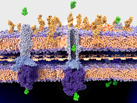
Free Images: "bestof:Gap cell junction-de.svg the diagram shows a cell union called Gap junction Darstellung einer Gap Junction Zell-Zell-Kanal Image Gap cell junction svg"
Gap cell junction-de.svg
Gap cell junction-cs.svg
Gap cell junction-es.svg
Gap cell junction-zh.svg
Gap cell junction-en.svg
Gap cell junction it.svg
Gap cell junction-zh-tw.svg
Gap cell junction keys.svg
Adherens Junctions structural proteins.svg
Adherens Junctions structural proteins-fr.svg
Cell junction simplified cs.svg
Cell junction simplified it.svg
Cell junction simplified en.svg
Cell junction simplified pt.svg
Gap cell junction-gl.svg
Cellular tight junction-fr.svg
Cellular tight junction-en.svg
Gap cell junction zh-cn.svg
Gap cell junction-uk.svg
Connexon and connexin structure cs.svg
Adherens Junctions structural proteins-LangNeutral.svg
Connexon and connexin structure.svg
Connexon and connexin structure fr.svg
Adherens Junctions structural proteins-ja.svg
Adherens Junctions structural proteins it.svg
Desmosome cell junction de.svg
Electromagnet with gap.svg
Bandgap-reference.svg
Cellular tight junction keys.svg
Cellular tight junction-sv.svg
ShockleyQueisserFullCurve (DE).svg
Second gap cell lifecycle.svg
First gap cell lifecycle.svg
Desmosome cell junction pl.svg
Desmosome cell junction gl.svg
Desmosome cell junction it.svg
Desmosome cell junction en.svg
Desmosome cell junction uk.svg
Cell junction simplified zh traditional.svg
桥粒(细胞连接).svg
Cellular tight junction-it.svg
ExampleOfBulkPhotovoltaicEffect.svg
Connexon and connexin structure gl.svg
ShockleyQueisserBreakdown.svg
Schottky barrier minority carrier injection.svg
Human genome by functions.svg
Acrosome reaction diagram de.svg
Cell junction simplified zh.svg
Cell junction simplified gl.svg
Cell junction simplified uk.svg
Cell membrane detailed diagram de.svg
Cell membrane detailed diagram blank.svg
Cell membrane detailed diagram en.svg
Complete neuron cell diagram de.svg
Cell membrane detailed diagram id.svg
U 2000 EN QUAD.svg
Animal cell structure pt.svg
Schema pflanzliche Zellwand.svg
Endomembrane system diagram cs.svg
Endomembrane system diagram hu.svg
Endomembrane system diagram nl.svg
Endomembrane system diagram ka.svg
Endomembrane system diagram ln.svg
Endomembrane system diagram zh.svg
ShockleyQueisserFullCurve.svg
Cellular tight junction-gl.svg
ShockleyQueisserZoomedIn.svg
Cellular tight junction-uk.svg
Endomembrane system diagram en.svg
Optical flat interference.svg
ShockleyQueisserVOC.svg
Coptic-Cross.svg
Right Ventricular hypertrophy.svg
Right Ventricular hypertrophy-IT.svg
Bluebaby syndrom.svg
Phylogenetic_tree-de.svg
ShockleyQueisserBreakdown2 (DE).svg
Straddling gap heterojunction band diagram.svg
Cell membrane detailed diagram es.svg
Osmotic pressure on blood cells diagram pl.svg
Osmotic pressure on blood cells diagram-es.svg
Osmotic pressure on blood cells diagram-ru.svg
Osmotic pressure on blood cells diagram (zh-cn).svg
Osmotic pressure on blood cells diagram-lv.svg
Prokaryote cell diagram pt.svg
Prokaryote cell diagram-bn.svg
Prokaryote cell diagram sr.svg
Cell membrane detailed diagram pt.svg
Prokaryote cell diagram he.svg
Cell membrane detailed diagram zh.svg
Diagram human cell nucleus sk.svg
Complete neuron cell diagram he.svg
Complete neuron cell diagram without text.svg
Diagram human cell nucleus (zh-cn).svg
Cell membrane detailed diagram lv.svg
Diagram cell nucleus no text.svg
Prokaryote cell diagram cs.svg
Diagram human cell nucleus cs.svg
Prokaryote cell diagram international.svg
Diagram human cell nucleus hu.svg
Terms of Use   Search of the Day