Click Here for More Images from iStock- 15% off with coupon 15FREEIMAGES 
Petri dish with bacteria colonies, isolated on white
Close-up shot of a petri dish for checking an antibiotic in the laboratory.
Bacterial culture plate examination by a female researcher in microbiology laboratory
Double zones can be seen around 3 antibiotic disks. This is a clear sign of more than one bacterium being present. To measure antibiotic susceptibility for each bacterium it will be necessary to isolate E. coli and Klebsiella on separate agar plates.
Set of Petri dishes with different culture samples on white background, top view
Different types in cell development glass dishes on desk. Group of Petri dishes with different culture media and micro organisms grown.
Hand with Petri dish or culture media with bacteria on white background with clipping, Test various germs, virus, Coronavirus, COVID-19, Microbial population count, Food science.
View of e.coli bacterium growing in petri dish seen through magnifying glass
E.coli bacterial growth or colonies on an Eosin Methylene Blue or EMB agar plate in a microbiology lab.
In vitro bacterial culture
Clean culture of aerobic bacteria on agar plate, biohazard sign in background. Selective focus on petri dish, slightly blue toned, AdobeRGB color profile. Content of my lightbox medical & sciences:
Scientist or doctor hand with a blue glove holding a petri dish with a selective culture of Escherichia coli bacteria
ESBL negative in sensitivity test in muller hinton media.
Top view of petri dish and culture media with bacteria on white background, solid media, nutrient agar, Test various germs, virus, Coronavirus, Corona, COVID-19, Microbial population count.
Candida grown on agar plate
Backgrounds of Characteristics and Different shaped Colony of Bacteria and Mold growing on agar plates from Soil samples for education in Microbiology laboratory.
In vitro bacterial culture
A listeria monocytogenes growing on an agar plate isolated on a black background
E. coli bacteria growing in laboratory dish inspected in a food laboratory
Fungal colonies isolated in the scientific research laboratory
Escherichia Coli (E. Coli.)
All drugs are resistance in sensitivity test.
Backlit composition on white background of different microbes growing on colture media
Antimicrobial susceptibility testing in petri dish. Microbiologist check Antibiotic resistance of bacteria.
Close-up shot of a petri dish for checking an antibiotic in the laboratory.
Petri dish or culture media with bacteria on white background with clipping, Test various germs, virus, Coronavirus, Corona, COVID-19, Microbial population count.
Liver Fluke Section under light microscope with white background
Differentiation of bacteria
stx2a is a subtype of Shiga toxin-producing E. coli (STEC) linked to Hemolytic Uremic Syndrome (HUS), a severe condition impacting kidney function and blood clotting. STEC poses a significant risk of foodborne illness and can spread within communities including family members.
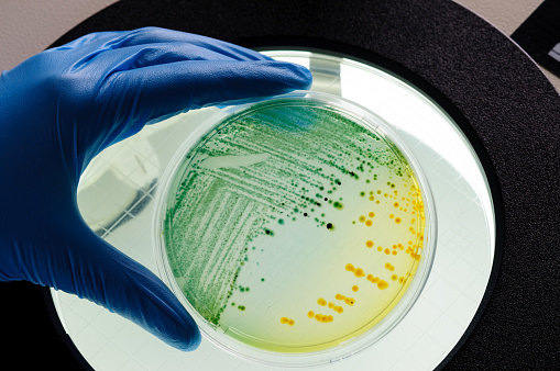
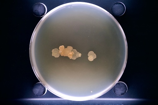
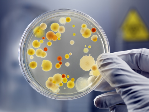
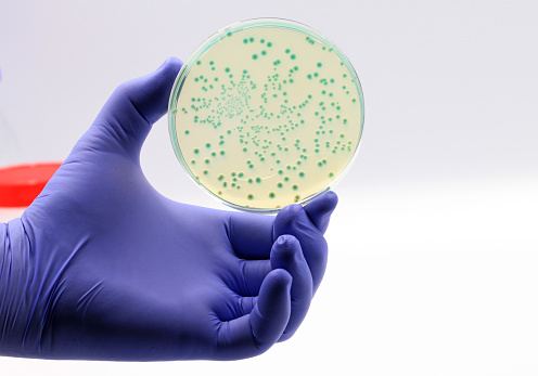
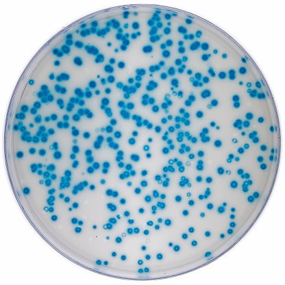
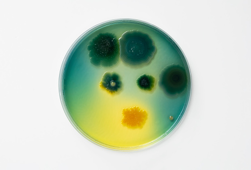
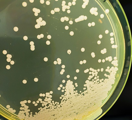
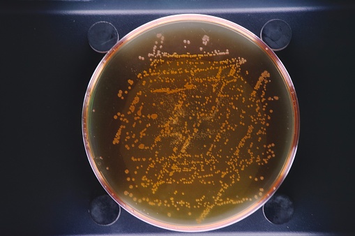
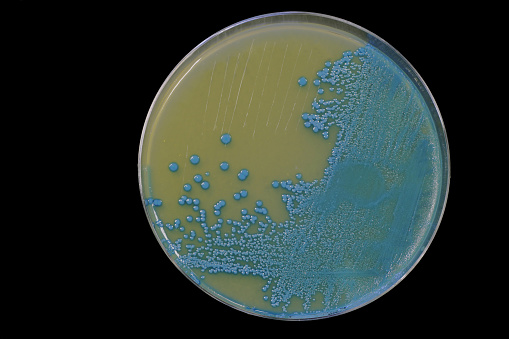
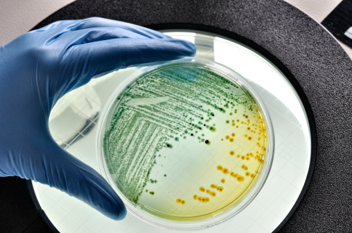
Free Images: "bestof:Fluorescent.jpg en plasmid pGLO in E coli DH5 alpha own 25kartika 2010-03 Petri dishes cultures Escherichia coli"
Konjugation-ecoli.svg
MinCDE System.svg
E. coli 2011 Map.svg
Konjugation-hfr-ecoli.svg
MinCDE System2.svg
Flagellum base diagram en.svg
Flagellum base diagram-fr.svg
QWeibull cdf.svg
QWeibull pdf.svg
Fluorescent.jpg
Macconkey e coli.jpg
Ecoli colonies.png
Sterione bild1.png
EColiUSDAfromPinaFratamico.jpg
EColiCRIS051-Fig2.jpg
Konjugation-ecoli.png
Asparaginase2 Ecoli 3ECA.png
ExonucleaseIIIfromEColi.png
E.coli operon regulation network.jpg
P-aeruginosa fluoreszenz.jpg
Sterione bild2.png
Ecoli metapopulation.jpg
OGDH-Ecoli-2JGD.png
Macconkey LF NLF.jpg
Bacillus sps various types blood agar.jpg
Vibrio cholerae on TCBS agar.jpg
Podospora anserina.jpg
Muller Hinton agar with MRSA.jpg
Adn bleu de toludine.JPG
MacConkey agar with LF and LF colonies.jpg
Lactose non lactose fermenters on CLED agar.jpg
Binder CB 210 incubator interior.jpg
E.coli image.jpg
Ciclo celular de Escherichia coli.jpg
Isocitrate dehydrogenase of Escherichia coli Complex.jpg
Diverse e Coli.png
Tsh-associated self-cleaving domain (Escherichia coli) and similar.jpg
Saureus.JPG
Wankomycyna.JPG
Fungiinperfecta2.JPG
EcKARI p-domain NADPH.png
Carboxytransferase 2F9Y E-Coli.png
BCCP.png
ClC-EC 3NMO.jpg
Biotin carboxylase rainbow.png
Porcine Mitochondrial IDH 1lwd.jpg
Coca-cola pl.jpg
Primase 3B39.png
Crystal structure.jpg
Sterilization_effects_of_negative_air_ionization.jpg
Bile-escul.JPG
P-aeruginosa pigment.jpg
E coli beta clamp 1MMI.png
Chicken fricassee.jpg
IMP allosteric binding site.JPG
Tinola.jpg
Gélose Chapman.JPG
Blue white assay Ecoli.jpg
Fluorescent bulb check 1.JPG
SutterlinAlsace1942.jpg
GE biax 2D.JPG
Fluorescent light bulb check 2.JPG
Mary_McLeod_Bethune_with_a_Line_of_Girls_from_the_School_WDL4013.png
Gower meat selection at Swansea restaurant.jpg
WNJN-TV Montclair dishes jeh.jpg
WNJN-TV Montclair signs jeh.jpg
Burgos - San Esteban, orfebreria 02.jpg
Figure-1.JPG
Tomyumsoup.png
Romanian_Schools_for_Aromanians_and_Meglenoromanians.JPG
Trollope Thomas.JPG
FRAD007_061_Le_diplôme_de_mérite_d’une_écolière_de_Melle_(Deux-Sèvres),_item_1.jpg
Terms of Use   Search of the Day