Click Here for More Images from iStock- 15% off with coupon 15FREEIMAGES 
After dopamine binding, the dopamine receptor changes his shape and binds the inactive G protein.The G protein expels its GDP molecule replacing it by GTP. and falls into two pieces. The Galpha subunit binds to and activates the enzyme adenylyl cyclase, which produces then cyclic AMP spreading a signal through the cell.
Cell Membrane Phospholipid Structure,Phospholipid bilayer of the cell membrane,Lipid bilayer
abstract binary random white background 3D renderingabstract background 3D rendering - letters conceptabstract background 3D rendering -  multi ethnic conceptabstract background 3D rendering - letters concept
Phospholipid bilayer membrane structures that make up living cells and their contents. This is an image of membrane proteins in the membrane.\nThis image, which looks like a plastic model, evokes biology and biochemistry. It can be used as a cover for magazines, textbooks and presentation materials.\nThis image was not created by AI.
3D cartoon and Gaussian surface models, chain id color scheme, PDB 7t3a, white background
Inside protein molecule
Biological protein and molecule, 3d rendering. 3D illustration.
Abs technology COVID-19 research - 3d rendered image. Pandemic wave.\nViral Infection concept. Delta, Omicron (B.1.1.529) variant. Sars-cov-2, 2019-nCoV, Coronavirus.\nAntibody, Antigen, stealth, Vaccine technology concept.
Bacterial multidrug efflux pumps (E-CmeB) expel antibiotics  from inside the bacterium (bottom): ciprofloxacin (yellow), chloramphenicol (violet),  ampicillin (red).
plant Pine Young Staminate Cone L.S. under light mircoscope with white background
microscopic view of plant cells for botanic education and analysis
Painted on Digital Tablet; Mosaic Pixelated, Abstract Planets
Proliferating cell nuclear antigen (PCNA) is a DNA clamp that acts as a processivity factor for DNA polymerase δ in eukaryotic cells and is essential for replication. PCNA is a homotrimer and achieves its processivity by encircling the DNA, where it acts as a scaffold to recruit proteins involved in DNA replication, DNA repair, chromatin remodeling and epigenetics. 3D cartoon and Gaussian surface models, chain instance color scheme, based on PDB 3wgw, white background.
Texture pattern on marble of stone surface of agate slice.
Lime trunk
Close up of cristal beads
Light photomicrograph of pine tree wood seen through a microscope
plant rape flower cross section under light mircoscope with white background
3D illustration of sanitary piping
corn stem micrograph with dye
Plant tissue Structure, section (tissue) of stem plant tissue under a light microscope.
A ball and stick model of a Polytetrafluoroethylene molecule, also known as PTFE or most commonly Teflon. It is a polymer consisting of only carbon and fluorine atoms. It is used as a non-stick coating for cookware, as a lubricant, and it is also used to contain highly reactive chemicals as it is extremely non-reactive.
The algae species Pediastrum boryanum under the microscope.
Chloroplast isolated on black
3D cartoon and Gaussian surface models, PDB 3hg3, white background
Slice of natural crystal for patterns and textures use with high resolution.
microscopic view of plant cells for botanic education and analysis
macro photo of different structures, hexagons, pentagons,  in green colored water soap bubble
Computer Motherboard Architect blueprint
Set gemstones crystal minerals for relaxation and meditation. Energy healing minerals. Witchcraft, Crystal Ritual, Relaxing Chakra, aura readings. Flat lay, top view
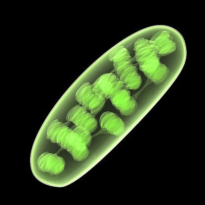
Free Images: "bestof:Euglenid pellicula scheme.svg Scheme of euglenid pellicula structure 1 cell membrane; 2 protein pellicular strip; 3 microtubules; 4 traverse filament; 5"
Euglenid pellicula scheme.svg
Scheme facilitated diffusion in cell membrane-zh (zh-tw).svg
Scheme facilitated diffusion in cell membrane-en.svg
Scheme facilitated diffusion in cell membrane-zh.svg
Scheme facilitated diffusion in cell membrane-eo.svg
Scheme facilitated diffusion in cell membrane-ca.svg
Lipid raft organisation scheme.svg
Scheme simple diffusion in cell membrane-es.svg
Scheme simple diffusion in cell membrane-de.svg
Scheme simple diffusion in cell membrane-zh (zh-cn).svg
Scheme simple diffusion in cell membrane-zh (zh-tw).svg
Scheme facilitated diffusion in cell membrane-es.svg
Scheme simple diffusion in cell membrane-en.svg
Scheme simple diffusion in cell membrane-de 02.svg
Scheme facilitated diffusion in cell membrane-fr.svg
Scheme simple diffusion in cell membrane-pl.svg
Scheme simple diffusion in cell membrane-fr.svg
Scheme simple diffusion in cell membrane-ca.svg
Scheme sodium-potassium pump-it.svg
Scheme sodium-potassium pump-en-2.svg
Scheme sodium-potassium pump-ja.svg
Scheme sodium-potassium pump-zh.svg
Scheme sodium-potassium pump-en.svg
Scheme sodium-potassium pump-pl.svg
Scheme sodium-potassium pump-bs.svg
Scheme sodium-potassium pump-he.svg
Scheme sodium-potassium pump-zh (zh-tw).svg
Scheme secundary active transport-en.svg
Scheme secundary active transport-heb.svg
Scheme sodium-potassium pump-el.svg
Scheme secundary active transport-pl.svg
Scheme sodium-potassium pump-cs.svg
Scheme sodium-potassium pump-de.svg
Scheme sodium-potassium pump-ca.svg
Cellular tight junction keys.svg
Scheme simple diffusion in cell membrane-he.png
Scheme facilitated diffusion in cell membrane-de.png
Scheme simple diffusion in cell membrane-de.png
3D_protein.jpg
GIRK 1N9P.png
Scheme sodium-potassium pump-ar-2.png
Chlamydomanas reinhardtii Flagella 9 - TEM.jpg
Terms of Use   Search of the Day