Click Here for More Images from iStock- 15% off with coupon 15FREEIMAGES 
A ball and stick model of Human Insulin. Isolated on black.
Bacterial multidrug efflux pumps (E-CmeB) expel antibiotics  from inside the bacterium (bottom): ciprofloxacin (yellow), chloramphenicol (violet),  ampicillin (red).
3D cartoon and Gaussian surface models, chain id color scheme, PDB 7t3a, white background
The three-dimensional structure of proteins is represented in a variety of conformations. This image is based on the ribbon structure of a protein. It is suitable for biology notebooks and book covers.\nThis image has not been created by AI.
Calcidiol molecule, also called calcifediol, major circulating metabolite of vitamin D3 produced in liver, 3D illustration
Cell Membrane Phospholipid Structure,Phospholipid bilayer of the cell membrane,Lipid bilayer
Zearalenone or ZEN or RAL is a potent estrogenic metabolite. 3d illustration
3d Rendering Molecular Structure,Ozone,Hydrogen and oxygen
Science background with molecule or atom
A model of a molecule of arginine, an amino acid. Amino acids are the building blocks of proteins and have many functions in metabolism. In particular, Arginine has an important function in the immune system, wound healing and cell division.
From left to right: the NMDA and  AMPA receptors transport calcium cations into neurons after being activated by the neurotransmitter glutamate and  the GABA receptor (right) transport chloride anions after the activation by gamma aminobutyrate. PDB entries: 6wht,  3kg2, 6d6u
Cells in the body examined by endoscope
Cell membrane cholesterol is shown in pink. 3D cartoon and Gaussian surface models, PDB 7rpk, white background.
3D Rendered Illustration, visualization of Neurons firing neurotransmitters in the synaptic gap.
Baryte or barite is a mineral consisting of barium sulfate. The baryte group consists of baryte, celestine, anglesite and anhydrite. 3d illustration
Computer generated abstract concept image of 3d spheres like metaphor of abstract business processes.
Molecular model of insulin hormone, 3D illustration. It is produced by pancreas and takes part in glucose and lipid metabolism
Molecular structure of Sulfur dioxide SO2
Horizontal banner with model of abstract molecular structure. Background of gray color with glass atom model. Copy space for your text. 3d render
Fictitious molecule model inside red and blue pill isolated on white.
3D cartoon and Gaussian surface models, chain id color scheme, PDB 6jod, putative membrane is shown, white background
A molecular model of the flavonoid Quercetin.  An antioxidant and anti-inflamatory, it is used as a nutritional suplement.  It is commonly found in many fruits and vegetables and is a significant chemical component of tea.  Isolated on white.
Squalane is a hydrocarbon derived by hydrogenation of squalene. 3d illustration
Liquid bubbles up Fluid Twirl
copper hydroxide, pools abstract structure
3D realistically coloured hexagonal shapes of connected spheres, background with copy space. Futuristic illustration design template for scientific presentation. Molecular business, science, teamwork concept.
The calcitonin gene related peptide (yellow) binds to its receptor (blue) on neurons and smooth muscle cells of cerebral blood vessels, activating a signal cascade through G-proteins (dark blue) in this cells that  leads to a dilatation of the blood vessels (vasodilatation).
Inside protein molecule
Prednisone is a glucocorticoid medication mostly used to suppress the immune system and decrease inflammation in conditions such as asthma, COPD, and rheumatologic diseases.
A molecular model of Bisphenol A or BPA.  It is a common building block in the production of many epoxy resins and plastics including polycarbonate plastic. As such it is present in a huge range of food packaging and household products. There are now concerns about its health effects, particularly its effect on children, infants and fetuses.   Isolated on white.
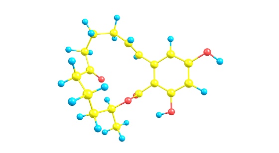
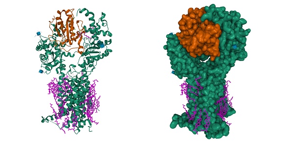
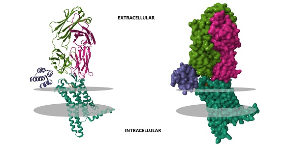
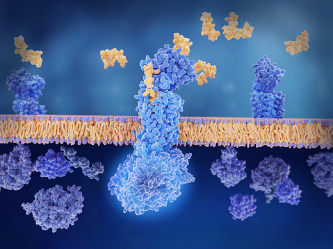
Free Images: "bestof:Estrogen receptor+lasofoxifene.png en A crystal structure of the ERalpha ligand-binding domain complexed with lasofoxifene pl Struktura receptora estrogenowego"
RDX1.svg
Nomex.svg
Dislocation.svg
Fikocyjanobilina 007.svg
Sulfurothioic S-acid.svg
Average prokaryote cell-pl.svg
Cryst struct deficit.svg
Main protein structure levels pl - secondary structure.svg
Beta-D-Glucopyranose.svg
2ph1 pdb gallery nucleotide binding-protein-2 (MinD homolog).svg
Estrogen receptor+lasofoxifene.png
RXRalpha-alitretinoin.png
Estrogen receptor beta.png
Steroid receptor.png
Lasofoxifene.png
The crystal structure of the GPIbα N-terminal domain depicting important ligand binding sites.png
Streptokinase 1BML.png
Crystal structure of modified Gramicidin S horizontally.png
CD4+MHCII 1JL4.png
Crystal Structure of the FokI endonuklease - PDB-2FOK.png
Crystal structure of the FokI endonuclease - PDB-2FOK 2.png
Crystal Structure of the Ku80 - 11.png
Crystal Structure of the Ku80 - 37.png
Crystal Structure of the Ku80 - 38.png
Crystal Structure of the Ku80 - 39.png
Crystal Structure of the Ku80 - 40.png
Crystal Structure of the Ku70 - 41.png
Crystal Structure of the Ku70 - 42.png
Crystal Structure of the Ku70 - 43.png
Crystal Structure of the Ku Heterodimer - 15.png
Crystal Structure of the Ku Heterodimer - 16.png
Crystal Structure of the Ku Heterodimer - 17.png
Crystal Structure of the Ku Heterodimer - 2.png
Crystal Structure of the Ku Heterodimer - 20.png
Crystal Structure of the Ku Heterodimer - 22.png
Crystal Structure of the Ku Heterodimer - 23.png
Crystal Structure of the Ku Heterodimer - 26.png
Crystal Structure of the Ku Heterodimer - 30.png
Crystal Structure of the Ku Heterodimer - 31.png
Crystal Structure of the Ku Heterodimer - 33.png
Crystal Structure of the Ku Heterodimer - 36.png
Crystal Structure of the Ku Heterodimer - 35.png
Crystal Structure of the FokI endonuclease - PDB-2FOK.png
Tal structure of FokI endonuclease -PDB-2FOK 3.png
1PWU.png
3EVA.png
1bda.png
Oxyluciferin-Luciferase.Micro.png
RORC 3L0L.png
Niklozamid.png
Pyrazinamidase.png
Human CES1 1mx1.jpg
Lasofoxifene structure.png
Bluelaser1a.jpg
Bluelaser1b.jpg
Orcine.png
Chain_A,_Crystal_Structure_Of_Mcpip1_Conserved_Domain_With_Zinc-finger_Motif.png
CBX1+EMSY 2FMM.png
1pn2.png
Doxepin.png
AIF factor.png
Bak protein.png
Bax protein.png
Bcl-2 protein.png
Struktura-blue1.jpg
Corticosteroid-Binding Globulin.png
Struktura-blue1.png
Furfuramide-3D-vdW.png
RAP1-rasmol.GIF
Terf2 and tin2.JPG
1FMK (c-Src).png
Bcr abl sti.jpg
Bunte-salts-structure.png
Struktura drzewostanow Puszczy Bialowieskiej.jpg
Perovskita-wiki.jpg
4f0a bio r 500.jpg
Carbon lattice diamond.png
Iceviistructure-ru.gif
Bcl-xl.jpg
Ribbon diagram depicting the crystal structure of the GPIbβ extracellular domain.png
Isosorbide dinitrate.png
Vts1P SRE Recognition SAM domain.jpg
HLA-A24.png
Miglustat-enzyme complex.png
Crystal structure of SopE.png
PDB-2RGT-Complex Lhx3 and Isl1.png
Aztreonam.png
LDLR Binding Module5 1ajj.PNG
Pepsin+Pepstatin 1PSO.png
Improving domain structure by Zureks.png
Interleukin-1b crystal stru.jpg
Interleukin-1a crystal str.jpg
1NBF-KGramatikoff.jpg
Kinesin motor domain 1BG2.png
CD86 structure.gif
Struktura płci i wieku w gminie Topólka (2009).tif
Mutated EGFR kinase domain in complex with gefitinib.png
Interleukin-1-receptor+IL-1beta 1ITB.png
PDB 3rd repeat of Reelin protein.jpg
Bovine_serum_albumin_3v03_crystal_structure.jpg
Terms of Use   Search of the Day