Click Here for More Images from iStock- 15% off with coupon 15FREEIMAGES 
Connective tissue, hyaline degeneration biopsy under light microscopy zoom in different areas
Host cells with spores (mold) are inside wood under the microscope for education.
Aerial view of loyster farm by Ardara, County Donegal - Ireland.
Blastomycosis is a fungal infection caused by inhaling Blastomyces dermatitidis spores. If it involves only the lungs, it is called pulmonary blastomycosis. Only about half of people with the disease have symptoms, which can include fever, cough, night sweats, muscle pains, weight loss, chest pain, and feeling tired.
Epithelial Cells in urine sediment.
Microscopic image of dog stomach cardiac region
Particles, medical concept
Lily leaf c.s. under microscope
Multicolored smears of face cosmetics on laboratory glass on a gray background. The concept of research and creation of cosmetics
Spinal Cord C.S. under light microscope with white background
Algae, a unicellular organism
Colorful seamless pattern with Crystal stones.
Prostate Cancer: Microscopic image of prostatic tissue, adenocarcinoma, malignant neoplasm, atypical epithelial cells.
Thymoma thymus.  Thymoma and thymic carcinoma are diseases in which malignant (cancer) cells form in the thymus. Thymoma is linked with myasthenia gravis and other autoimmune paraneoplastic diseases.
Cartilage cells under the microscope view.
Close-up shot of a petri dish for checking an antibiotic in the laboratory.
Stratified flat epithelium under microscope
plant Zea Stem C.S. under light mircoscope with white background
The study of plant tissues under the microscope in the laboratory.
Texture pattern on marble of stone surface of agate slice.
Bladder Cancer: High grade transitional cell carcinoma (TCC), Urothelial carcinoma
Photomicrograph of two rotifers. Rapidly rotating cilia at top of heads, internal organs visible. Live specimen. Wet mount, 10X objective, transmitted brightfield illumination.
Abstract foamy water liquid painted in green and pink color on stone background. Flat lay cocept.
Lymphocytic myocarditis. Myocarditis is an inflammation of the heart muscle. Lymphocytic refers to the type of cell seen in the affected tissue. A lymphocyte is a type of immune function cell.
Microscope photo of a sambucus stem with parenchyma cells.
Quantum Gravity 3d rendered image of a comprehensive exploration of Quantum Gravity theories. String theory, loop quantum gravity (LOG), causal dynamical triangulation, twistor theory, gravitational waves, spacetime, gravity waves, quantum mechanics, quantum entanglement concepts.
Histology microscope image of smooth muscle tissue (400x)
Close-up shooting of a green agate stone mineral surface isolated on white background. Its a macro shot shows nature beautiful details with back lit. Agate is a kind of natural gemstone. Abstract pattern. Good for Wallpaper texture background. Purple
Cross sections of the plant root under the microscope view for education
stx2a is a subtype of Shiga toxin-producing E. coli (STEC) linked to Hemolytic Uremic Syndrome (HUS), a severe condition impacting kidney function and blood clotting. STEC poses a significant risk of foodborne illness and can spread within communities including family members.
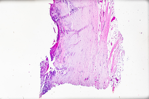
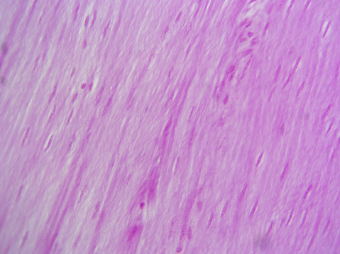
Free Images: "bestof:Epidermis-delimited.JPG This is a hematoxylin and eosin stained slide at 10x of normal epidermis Normal_Epidermis_and_Dermis_with_Intradermal_Nevus_10x JPG"
Bombus_terricola,_male,_face_2012-07-12-16.54.jpg
Epidermis-delimited.JPG
Normal Epidermis and Dermis with Intradermal Nevus 10x-cropped.JPG
Normal Epidermis and Dermis with Intradermal Nevus 10x.JPG
Base of Pilosebaceous Unit 10x.JPG
Insertion of sebaceous glands into hair shaft x10.jpg
Burkitt's_lymphoma_Histopathology.jpg
Trichinella_rat_BAM2.jpg
UIPlungbiopsy.jpg
Breast cancer cells (2).jpg
Breast cancer cells (1).jpg
Breast cancer cells.jpg
Terms of Use   Search of the Day