Click Here for More Images from iStock- 15% off with coupon 15FREEIMAGES 
Excretion is the process by which waste products of metabolism and other non-useful materials are eliminated from an organism.
plant Zea Stem C.S. under light mircoscope with white background
Striped mosquitoes life circle with white background. Life cycle of mosquitos isolate on white background.
Computational tomography image of the abdominal and pelvic regions.
forming of underskin cellulite illustration
Photomicrograph of filamentous blue-green algae, Oscillatoria species. Each band along the stand is one cell. This filament is very short probably having recently parted from a longer one. Filaments are typically many times longer. This algae is mobile, it moves and twists through the water. San Francisco Bay, California, USA. Live specimen. Wet mount, 40X objective, transmitted brightfield illumination. Note - motion blur of live specimen, very shallow depth of field, chromatic aberration and uneven focus are inherent in light microscopy.
question, learn, try, fail, succeed, share and grow - perseverance cycle, a sketch on a napkin, business, education and personal development concept
ERP - Enterprise Resource Planning
Microalgae under microscopic view, green algae, cyanobacteria, phytoplankton, diatom, algae mix collage background
Liver Fluke Section under light microscope with white background
Eggs of Hookworm in stool, analyze by microscope
The three-dimensional structure of proteins is represented in a variety of conformations. This image is based on the ribbon structure of a protein. It is suitable for biology notebooks and book covers.\nThis image has not been created by AI.
3D cartoon and Gaussian surface models, PDB 6gu2, white background
What micro plastic is made of - Chemical ingredients.\nClose up of microplastic on finger concept of water pollution and global warming
Chaetoceros is the largest genus of marine planktonic diatoms, phytoplankton
Trematode parasite under a microscope
Acetylcholine receptors in the lipid layer of cell membrane.
Abstract Globe World Maps On White
brown, beige and white fat cells,  adipocyte and lipocyte, cholesterol in a cells, adipose tissue, lipid droplet, \nfat in body, Obesity, Types of lipocytes dermis and hypodermis, nucleus, 3d render
Abstract technological design of an artificial molecular structure in many colors. Horizontal wide composition.
Mechanical properties of springs according to Hooke's law. Digital illustration.
Photomicrograph of rotifer, probably Rotaria rotatoria. Rapidly rotating cilia at top of head, internal organs visible. Green circles are algae. Live specimen. Original image at 40X, wet mount, transmitted brightfield illumination. Indistinct image due to motion blur of live specimen and very shallow depth of field of brightfield illumination.
MRI of the prostate gland reveals Focal abnormal SI lesion at left PZpl at apex as described; PI-RADS category 4, clinically\nsignificant cancer is likely.
Chloroplast isolated on black
Ultrasound examination of the abdominal cavity.
Close up of microplastic on female finger concept of water plastic pollution and global warming with text explaining the ingredients of micro plastic
eggs of parasite on white background.
Squash preparation of  onion root tip cells stained to reveal chromosomes,  Bright field illumination, light microscopy.  This image was taken with the X40 objective lens.  Depth of focus is extremely low at this magnification.  Smaller apertures give greater depth of focus, but image quality deteriorates and resolution is greatly reduced.  The aperture in this image was optimised for contrast and resolution. Chromatic abberation is inevitable in light microscopy, but has been kept to a minimum in this image.
3D cartoon and Gaussian surface models with a putative membrane shown, PDB 4p02, white background
innovation and technology
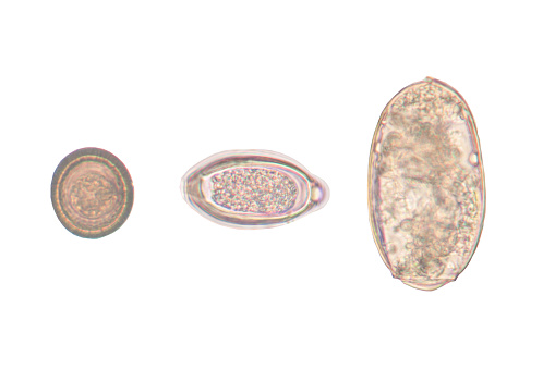
Free Images: "bestof:Enterobius vermicularis LifeCycle B.svg Enterobiasis Enterobius vermicularis<br/>Life cycle of Enterobius vermicularis<br/>Eggs are deposited on perianal folds"
Enterobius_vermicularis_LifeCycle_B.svg
Enterobius_vermicularis_LifeCycle_fr-version.svg
Enterobius_vermicularis_LifeCycle_pt-version.svg
Enterobius_vermicularis_LifeCycle.gif
Enterobius vermicularis LifeCycle-gl.gif
Enterobius vermicularis LifeCycle gl.gif
Enterobius_vermicularis_LifeCycle_cs.GIF
Eggs_of_Enterobius_vermicularis_5229_lores.jpg
Owsica.GIF
Head_of_Enterobius_vermicularis_human_pinworm_5230_lores.jpg
Enterobius_cellulosetape.jpg
Evermicularis_SC_posterior.jpg
Evermicularis_egg_UVa.jpg
Evermicularis_egg_UVb.jpg
E_vermicularis_eggs_tissue2.jpg
Evermicularis_egg_HBa.jpg
Evermicularis_egg_wtmt.jpg
E_vermicularis_eggs_tissue1.jpg
Evermicularis_SC_egg.jpg
Evermicularis_worm4_HB.jpg
Evermicularis_SC_anterior.jpg
Enterobius_vermicularis.jpg
Enterobius_vermicularis.png
Enterobius_vermicularis.jpg
Oxyuris_vermicularis.jpg
Threadworm.jpg
Oxyuris_vermicularis_female.jpg
Taxones_parasitarios.jpg
E._vermicularis_egg.png
Evermicularis_worm5_HB.jpg
Terms of Use   Search of the Day