Click Here for More Images from iStock- 15% off with coupon 15FREEIMAGES 
Anatomically accurate human male skull with colorized bones lateral or profile view isolated on white background with copy space 3D rendering illustration. Blank anatomical and medical chart.
Film x-ray human's pelvis
X-ray skull(oblique) of thai people
in Villajoyosa (Vila Joiosa), Spain
The posterior sacroiliac ligament is a compound ligament composed of three distinct bands 3d illustration
Digital medical illustration: Posterior (back) view (orthogonal) of human skull (includes teeth) and neck featuring vertebrae, discs and ligaments (includes visible Interspinale, Nuchae, Anulus, Flavum, Capsula and Intertransversarium).
CT SCAN of Chest and Abdomen Coronal and sagittal view with injection contrast media for diagnosis chest and abdomen disease.
Magnetic resonance images of the brain (MRI brain). Computed resonance imaging of the human brain.
Anatomy of the pelvis bones, including the ilium, ischium, sacrum, and pubis, photorealistic 3D illustration. Front view. Perfect for educational or medical purposes.
historic churches within the historic city, Zadar, Croatia,
skull xray isolated on white background
X-ray of female hips, pelvis and spine
An EXCELLENT medical scan taken by a radiologist of a horse's right hoof; imagery was required to make proper diagnosis and determine the extent of the mare's injuries.
Basilica of Santa Croce, a church with great historical and architectural heritage that was once a synagogue. Cagliari. Sardinia. Italy.
Panoramic and 3D dental x-ray many shots
Cogs marks on a spray-painted surface.
Human Skull X-ray images AP and Lateral View isolated on Black Background
Santa Maria della Scala, Siena, Tuscany, Italy
The sacrum is the triangular bone just below the lumbar vertebrae 3d illustration
Magnetic resonance images of the brain (MRI brain). Computed resonance imaging of the human brain.
CT scan of a patient with missing chewing tooth and malocclusion.
CAT scan of human abdomen...man with aneurysm of abdominal aorta
Drawn and measured very precisely by Antoine Desgodetz Architect in 1676.
Human skull X-ray film
normal head  scan brain window
coat of arms of the city of Lille in Northern France on the facade of an old building; Lille, France
Very good quality X-ray image of normal human skull front (AP) view and side (Lateral) view, Process in blue tone isolated on black background.
The Great Clock or Gros Horloge in Rouen, Normandy
3D rendering image of Face , Facial bone ,skull front and both side view .clipping path
Front door of church
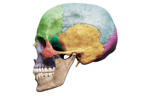
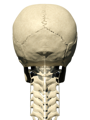
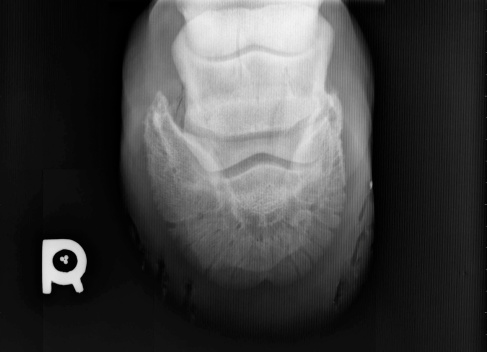
Free Images: "bestof:Dmanisi D-2282.jpg Dmanisi skull D-2282 reconstructed The cranium vault retains much of the face and a fragment of the maxilla but has post-mortem deformation"
Dmanisi D-2282.jpg
Dmanisi D-2282 left.jpg
Terms of Use   Search of the Day