Click Here for More Images from iStock- 15% off with coupon 15FREEIMAGES 
Microscopic image of Microscopic image of nymphaea of aqustio stem
Showing Light micrograph Type of Tissue Human under the microscope in Lab.
Illustration of animal cell anatomy
3d cell organelle - nucleus (Chromatin,  Rough Endoplasmic Reticulum, Nucleus Shell, Nucleoplasm, Nucleolus)
Cotton plant root, under the light microscope. Longitudinal section through the root of Gossypium hirsutum, upland or also Mexican cotton. Light micrograph at 20X magnification. Isolated, over white.
High quality 3d render of cells
Nephroblastoma with heterologous elements (teratoid Wilms' tumor). Wilms tumor (also called Wilms' tumor or nephroblastoma) is a type of childhood cancer that starts in the kidneys. It is the most common type of kidney cancer in children.
Components of Eukaryotic cell, nucleus and organelles and plasma membrane - 3d illustration
Viral Infection Monkeypox Virus - 3d rendered image. Abstract biomedical illustration. \nAntibody, Antigen, Vaccine technology concept.
The study of plant tissues under the microscope in the laboratory.
Cells on scientific background.
plant Zea Stem C.S. under light mircoscope with white background
Highly detailed image of human egg cell isolated on white background.
Natural agate surfaces, suitable for backgrounds and wallpapers.
Motor Neuron cells under the microscope
Epithelioid mesothelioma is a cancer subtype caused by asbestos. It is the most common mesothelioma cell type, accounting for 50% to 70% of cases.
3d rendering of biological animal cell with organelles cross section isolated on white. Animal cell with placed text annotations to all organelles
Viral Infection Monkeypox Virus - 3d rendered image. Abstract biomedical illustration. \nAntibody, Antigen, Vaccine technology concept.
Histological Pancreas human, Liver human, Vermiform appendix human and Kidney Human under the microscope for education.
Photomicrograph of flatworm,
Uterus with adnexa (biopsy): Cervix show chronic inflamatory cells, wall show secretory phase in endometrium and fallopian tube show paratubal cyst. fimbrial cyst, Paraovarian cyst.
Microscopic image of Microscopic image of nymphaea of aqustio stem
Organic sphere with chain structure inside, 3d rendering. Computer digital drawing.
digital illustration on the subject of science, medicine and biology
Epidermis. Electron microscope micrograph showing a keratinocyte of spinous layer. The epithelial cell has a polygonal shape, central nucleus with nucleolus, cytoplasm full of keratin filament bundles, and numerous dark desmosomes crossing the intercellular spaces.
plant rape flower cross section under light mircoscope with white background
High resolution 3d render of cells
Fatty liver disease (steatosis) is a common condition caused by having too much fat build up in your liver. A healthy liver contains a small amount of fat. It becomes a problem when fat reaches 5% to 10% of your liver's weight.
Leiden leaf c.s. under microscope
Slice of natural crystal for patterns and textures use with high resolution.
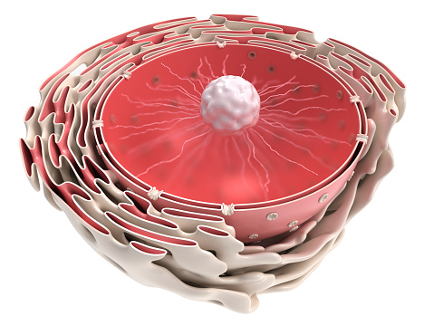
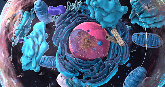
Free Images: "bestof:Diagram human cell nucleus sk.svg en A comprensive diagram of a human cell nucleous sk Schéma bunkového jadra Image Diagram human cell nucleus svg Translated"
Diagram human cell nucleus sk.svg
Diagram human cell nucleus cs.svg
Diagram human cell nucleus hu.svg
Diagram human cell nucleus id.svg
Diagram human cell nucleus sl.svg
Diagram human cell nucleus pt.svg
Diagram human cell nucleus pl.svg
Diagram human cell nucleus (zh-cn).svg
Diagram human cell nucleus tr.svg
Diagram human cell nucleus.svg
Diagram human cell nucleus fr.svg
Diagram human cell nucleus numbered version.svg
Diagram human cell nucleus oc.svg
Diagram cell nucleus no text.svg
Diagram human cell nucleus ita.svg
Diagram human cell nucleus ja.svg
Diagram human cell nucleus es.svg
Diagram human cell nucleus uk.svg
Complete neuron cell diagram en.svg
Complete neuron cell diagram id.svg
Complete neuron cell diagram fr.svg
Complete neuron cell diagram es.svg
Complete neuron cell diagram de.svg
Complete neuron cell diagram ca.svg
Complete neuron cell diagram numbered large numbers.svg
Complete neuron cell diagram without text.svg
Complete neuron cell diagram it.svg
Prokaryote cell diagram cs.svg
Schematic diagram of the human eye sk.svg
Gray5_nl.svg
Plant cell wall diagram mk.svg
Gray5_de.svg
Complete neuron cell diagram pl.svg
Complete neuron cell diagram ru.svg
Complete neuron cell diagram numbered.svg
Gap cell junction it.svg
Average prokaryote cell- lt.svg
Gray5.svg
Gray5-ja.svg
Gray5_-_es.svg
Prokaryote cell diagram he.svg
Cell membrane detailed diagram id.svg
Complete neuron cell diagram zh.svg
Complete neuron cell diagram zh-hant.svg
Complete neuron cell diagram pt.svg
PEM fuel cell.svg
Stomach-fr.svg
Human skeleton front sl.svg
Human skeleton back sl.svg
Human skeleton back vi.svg
Schema pflanzliche Zellwand.svg
Animal cell structure pt.svg
Cell junction simplified pt.svg
Zelula prokarioto tipikoa.svg
Prokaryote cell-ro.svg
Average prokaryote cell cs.svg
Cell junction simplified it.svg
Animal cell structure de.svg
Average prokaryote cell- fr.svg
Average prokaryote cell- nl.svg
Endomembrane system diagram nl.svg
Endomembrane system diagram ln.svg
Endomembrane system diagram cs.svg
Human skeleton front pl.svg
Human skeleton back pl.svg
Digestive system diagram an.svg
Human skull side suturas pl.svg
Human skull front bones pl.svg
Digestive system diagram sr.svg
Digestive system diagram lv.svg
Human skull side details pl.svg
Digestive system diagram es.svg
Digestive system diagram mk.svg
Human skull side details right pl.svg
Animal cell structure sv.svg
Digestive system diagram no labels arrows.svg
Flagellum base diagram hu.svg
Solid oxide fuel cell protonic mk.svg
Stomach colon rectum diagram-ar.svg
Stomach colon rectum diagram-is.svg
Stomach colon rectum diagram-th.svg
Thyroid system ko.svg
Turgor pressure on plant cells diagram (zh-tw).svg
Stomach colon rectum diagram-fr.svg
Clitoris anatomy labeled-en no.svg
Human skull side complete pl.svg
Human genome by functions.svg
Animal cell structure id.svg
Plant cell wall diagram-en.svg
Animal mitochondrion diagram id.svg
Animal mitochondrion diagram zh tw.svg
Average prokaryote cell- id.svg
Brain_diagram_ja.svg
Animal mitochondrion diagram zh hk.svg
Schematic diagram of the human eye he.svg
Cell membrane detailed diagram pt.svg
Schematic diagram of the human eye ta.svg
Animal mitochondrion diagram ml.svg
Animal mitochondrion diagram zh ml.svg
Cell membrane detailed diagram pl.svg
Terms of Use   Search of the Day