Click Here for More Images from iStock- 15% off with coupon 15FREEIMAGES 
3d illustration of clostridium bacteria
Particles, medical concept
Microscopic photo of a thin section of calcareous tufa of Holocene age.
Photomicrograph of filamentous blue-green algae, Oscillatoria species. Each band along the stand is one cell. This filament is very short probably having recently parted from a longer one. Filaments are typically many times longer. This algae is mobile, it moves and twists through the water. San Francisco Bay, California, USA. Live specimen. Wet mount, 40X objective, transmitted brightfield illumination. Note - motion blur of live specimen, very shallow depth of field, chromatic aberration and uneven focus are inherent in light microscopy.
Epidermis. Electron microscope micrograph showing a keratinocyte of spinous layer. The epithelial cell has a polygonal shape, central nucleus with nucleolus, cytoplasm full of keratin filament bundles, and numerous dark desmosomes crossing the intercellular spaces.
Bone marrow plasma cells .Plasma cell neoplasms are diseases in which the body makes too many plasma cells. Plasma cell neoplasms can be benign or malignant. Diagnosed with Immunostain CD138 positive.
Freshwater Closterium algae (unicellular charophyte green algae) - optical microscope x200 magnification
Cheyletiella blakei Mite - Small Cat pest view under a microscope
Euglena is a genus of single cell flagellate eukaryotes under microscopic view for study.
Abundant meningococci bacteria observed, with a notably absence of white blood cells in the spinal fluid from a critically ill meningitis patient. Meningococcal meningitis is the term used to describe a bacterial form of meningitis caused by Neisseria meningitidis. This form of meningitis is associated with high morbidity and mortality. \nSometimes the bacteria spread to people who have had close or lengthy contact with a patient with meningococcal disease. Those at increased risk of getting sick include: People in the same household, roommates and anyone with direct contact with the patient’s oral secretions, such as a kissing partner
Algae, a unicellular organism
plant Pine Cone-Female L.S. under light mircoscope with white background
Green algae cells background
Different germs in the human intestines called microbiome,Bacteria Lactobacillus in human intestine,Beneficial healthy intestinal bacterium microflora,Gut bacteria
Microscopic Rotifer or Rotifera under a microscope, freshwater bentic organism filtering water.
Pheochromocytoma, Site: Adrenal gland
Rhizopus (bread mold) is a genus of common saprophytic fungi,Rhizopus (bread mold) under the microscope.
Natural agate surfaces, suitable for backgrounds and wallpapers.
Licmophora sp. algae, marine and freshwater diatom under microscopic view. Genus of benthic, photosynthetic and epiphyte diatom
Photomicrograph of a freshwater clustering single-celled green algae,
Micrograph of a moss leaf, Dicranum, with elongated cells, and an elongated midrib (costa) that may be the water conducting cells of the leaf, with polarization at 200x.
Microscopic image of dog stomach cardiac region
Aerial view of loyster farm by Ardara, County Donegal - Ireland.
Blastomycosis is a fungal infection caused by inhaling Blastomyces dermatitidis spores. If it involves only the lungs, it is called pulmonary blastomycosis. Only about half of people with the disease have symptoms, which can include fever, cough, night sweats, muscle pains, weight loss, chest pain, and feeling tired.
centropyxis aculeata (amoeboid organism) under the microscope - optical microscope x400 magnification
Quantum Gravity 3d rendered image of a comprehensive exploration of Quantum Gravity theories. String theory, loop quantum gravity (LOG), causal dynamical triangulation, twistor theory, gravitational waves, spacetime, gravity waves, quantum mechanics, quantum entanglement concepts.
background colored macro bubbles of water
Liver Fluke Section under light microscope with white background
Human parasitic amoeba with pseudopodia, 3D illustration
Abstract particles
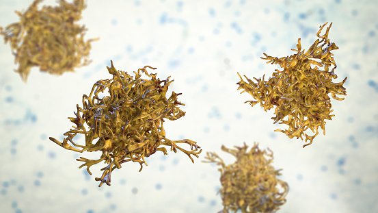
Free Images: "bestof:Clostridium difficile EM.png Obtained after an outbreak this micrograph depicts Gram-positive Clostridium difficile bacteria These C difficile organisms were"
Clostridium_difficile_EM.png
Protein tcdB pfam domains.png
Undiscovered species chart.png
Micrococcus_luteus_9758.jpeg
Tacuin_Ricotta31.jpg
Wall_Hanging_WDL2485.png
Johnson_Papyrus_WDL3959.png
Landscape_and_Notes_on_Shading_and_Perspective_WDL11283.png
Selected_Anecdotes_about_Su_Shi_and_Mi_Fu_WDL4693.jpg
Map_of_the_Belgian_Congo_WDL59.png
The_Battle_of_the_Monongahela_WDL9580.png
Kazuo_and_Masuko_Kamiya_as_Children_WDL4038.png
Layla, Issue 7, April 15, 1924 WDL2845.jpg
Layla, Issue 8, May 15, 1924 WDL2846.jpg
Layla, Issue 9, June 15, 1924 WDL2857.jpg
Layla, Issue 10, July 15, 1924 WDL2858.jpg
Layla, Issue 2, December 1924 WDL2860.jpg
Layla, Issue 3, January 1925 WDL2861.jpg
Layla, Issue 4, February 1925 WDL2862.jpg
Layla, Issue 5, March 1925 WDL2865.jpg
Layla, Issue 6, April 1925 WDL2866.jpg
Layla, Issue 7, May 1925 WDL2868.jpg
Layla, Issue 8, June 1925 WDL2869.jpg
Layla, Issue 9, July 1925 WDL3032.jpg
Layla, Issue 10, August 1925 WDL3033.jpg
Layla, Issue 2, November 15, 1923 WDL3055.jpg
Layla, Issue 3, December 15, 1923 WDL3056.jpg
Layla, Issue 4, January 15, 1924 WDL342.jpg
Layla, Issue 5, February 15, 1924 WDL452.jpg
Terms of Use   Search of the Day