Click Here for More Images from iStock- 15% off with coupon 15FREEIMAGES 
Squash preparation of plant cells stained to reveal chromosomes, various stages of mitosis are visible. Bright field illumination, light microscopy. This image was taken with the X40 objective lens. Depth of focus is extremely low at this magnification. Smaller apertures give greater depth of focus, but image quality deteriorates and resolution is greatly reduced. The aperture in this image was optimised for contrast and resolution. Chromatic aberration is inevitable in light microscopy, but has been kept to a minimum in this image.
3d rendering of Human cell or Embryonic stem cell microscope background.
vertical close up of Woman with shingles on the skin she feels very painful
Internal structure of an animal cell, 3d rendering. Section view. Computer digital drawing.
centropyxis aculeata (amoeboid organism) under the microscope - optical microscope x400 magnification
Photomicrograph of filamentous blue-green algae, Oscillatoria species. Each band along the stand is one cell. This filament is very short probably having recently parted from a longer one. Filaments are typically many times longer. This algae is mobile, it moves and twists through the water. San Francisco Bay, California, USA. Live specimen. Wet mount, 40X objective, transmitted brightfield illumination. Note - motion blur of live specimen, very shallow depth of field, chromatic aberration and uneven focus are inherent in light microscopy.
Microalgae under microscopic view, green algae, cyanobacteria, phytoplankton, diatom, algae mix collage background
Blastomycosis is a fungal infection caused by inhaling Blastomyces dermatitidis spores. If it involves only the lungs, it is called pulmonary blastomycosis. Only about half of people with the disease have symptoms, which can include fever, cough, night sweats, muscle pains, weight loss, chest pain, and feeling tired.
Bacteria of different shapes, cocci and rod-shaped bacilli, 3D illustration in old book vintage style
plant Zea Stem C.S. under light mircoscope with white background
Microscopic image of onion root tip cells undergoing mitosis. Anaphases and metaphases. 1000x magnified
Eczema
Squash preparation of stained onion root tip cells showing various stages of mitosis. Differential interference contrast optics on a Leica microscope, Image taken with the X40 objective lens.  Depth of focus is extremely shallow (just a few microns).  Differential interference contrast results in an optical slice horizontally through the specimen providing maximum possible resolution at this magnification and good contrast. Chromatic abberation is inevitable in light microscopy, but has been kept to a minimum in this image.
Freshwater Closterium algae (unicellular charophyte green algae) - optical microscope x200 magnification
Green algae cells background
Diffuse large B-cell lymphoma, or DLBCL, is a cancer that starts in white blood cells called lymphocytes.
Mitosis process, division of cell. Isolated on black background. 3d render
Liver Fluke Section under light microscope with white background
Background of mold in the wood of the house.
corn stem micrograph with dye
Epidermis. Electron microscope micrograph showing a keratinocyte of spinous layer. The epithelial cell has a polygonal shape, central nucleus with nucleolus, cytoplasm full of keratin filament bundles, and numerous dark desmosomes crossing the intercellular spaces.
Squash preparation of  onion root tip cells stained to reveal chromosomes,  Bright field illumination, light microscopy.  This image was taken with the X40 objective lens.  Depth of focus is extremely low at this magnification.  Smaller apertures give greater depth of focus, but image quality deteriorates and resolution is greatly reduced.  The aperture in this image was optimised for contrast and resolution. Chromatic abberation is inevitable in light microscopy, but has been kept to a minimum in this image.
This is a photomicrograph of cucumber pollen magnified 100 times.
Abs Bubonic plague bacteria - 3d rendered image structure view on black background. \nPlague Infection concept.  Hologram bacteria view.
Enlarge microscopic view of larvae stage of crustacean
plant rape flower cross section under light mircoscope with white background
Micrasterias is a unicellular green algae under the microscope view, phytoplankton
Myxoid liposarcoma.  Myxoid/round cell liposarcoma, or MRCLS, is one of several types of liposarcoma. Liposarcoma is a rare cancer that grows in the cells that store fat in the body. MRCLS usually grows in the arms and legs. These tumors grow slowly, and they can spread to other parts of the body
microscopy micrograph animal, conjugation of Paramecium caudatum, magnification 200X
Portraits of a beautiful young Argentinian woman - Buenos Aires - Argentina
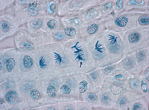
Free Images: "bestof:Citocinesis de mitosis eucariota.svg La mitosis es el proceso por el cual una célula eucariota se separa File Cytokinesis eukaryotic mitosis svg 2009-02-01 15"
Citocinesis de mitosis eucariota.svg
Tipos de exocitosis.svg
Endosimbiosis y primer cloroplasto.svg
Tipos de endocitosis.svg
Wrist and hand deeper palmar dissection-numbers.svg
Ciclo del nitrógeno 1.svg
Escudo Navalcarnero.svg
Escudo de Navalcarnero 2.svg
Ciclo del nitrógeno - Mudo.svg
Escudo de Navalcarnero 3.svg
Ciclo de oxígeno.svg
Universidad_de_Carabobo.svg
Ciclo de oxígeno - Mudo.svg
Mexico COA large.svg
Teulada.svg
Facultad_de_Ingeniería.svg
Coronas murales.svg
Escudo Nueva Esparta.svg
Jerez de los Caballeros.svg
Coat of arms of Nueva Esparta State.svg
Coat of arms of Panama.svg
Rainbow1.svg
Flag of Bellpuig.svg
Òdena Spain.svg
Flag of Thailand.svg
Coat of arms of Panama (green background).svg
Ogino-rueda.svg
Escudo de Asturias.svg
Escudo de Santa Cruz de Tenerife.svg
Álvaro Obregón, México DF.svg
Flag of Palafolls.svg
Escudo de Castrillón.svg
Coat of arms of Teulada.svg
Naval Jack of Spain.svg
Bandera de Barberà del Vallès.svg
Flag of Avià.svg
Swine flu infection exponent by county June 2009.svg
Flag of Sant Pau de Segúries.svg
Flag of Ivars d'Urgell.svg
Swine flu infection exponent by county FluTracker June 2009.svg
Flag of Europe.svg
Marcado watermarking.svg
Escudo de Torrevieja.svg
Cell membrane detailed diagram es.svg
Catalisis enzimatica1.svg
Coat of arms of Chile (c).svg
Escudo de Ávila.svg
Coat of arms of Chile 3D.svg
Coat of arms of Chile.svg
Fregenal de la Sierra.svg
Flower morphology - Flower longitudinal section bilabial Bignoniaceae Tecoma stans.svg
Cavendish Torsion Balance Diagram-es.svg
Escut de Balears.svg
Escut de Sant Adrià de Besòs.svg
Bandera de Almería.svg
Escudo de Andalucía (oficial2).svg
Partido Nacional Chile.svg
Escudo de Zamora.svg
Bandera de Almócita.svg
Demografía en Cádiz.svg
Flag of Aratoca.svg
Flag of Briceño (Boyacá).svg
Flag of Argentina and South Georgia and the South Sandwich Islands.jpg
Flag of Avinyó.svg
Escudo de la provicia de Soria.svg
Escudo de San Miguel de Allende.svg
Escudo de San Gil.svg
Escudo de Playas de Rosarito.svg
Phase space of cubic potential.svg
Notable mutations-es.svg
2011 Drug Harms Rankings es.svg
Flag of Paraguay (1811).PNG
Escudo de Murcia.svg
Azcapotzalco, México DF.svg
Escudo de Cenlle.svg
Coat of Arms of the Val d'Aran.svg
Flag of Sabanalarga (Antioquia).svg
Escut de Girona.svg
Escut de Bunyol.svg
Uchile.svg
E.L.M. de Guadiana del Caudillo (Badajoz).svg
Cicle de l'oxigen.svg
Bandera de Capdesaso-Huesca.svg
Flag of El Papiol.svg
Escudo de la Región de Murcia.svg
Escudo de armas de Santa Cruz de Tenerife.svg
Cytokinesis eukaryotic mitosis.svg
Flag of Constantí.svg
Bandera de El Paso.svg
IFT particle chlamydomonas.svg
Bandera de La Guardia de Jaén.svg
Seal of Tampa, Florida.svg
Escut d'Emperador.svg
Escudo de la Provincia de Alicante.svg
Escut de Godella.svg
Escudo de armas de Torrevieja.svg
Gernika-Lumo flag-2.svg
Escudo de la provincia de Cádiz.svg
Flag of Vilanova del Camí.svg
Elementary-particle-interactions-es.svg
Terms of Use   Search of the Day