Click Here for More Images from iStock- 15% off with coupon 15FREEIMAGES 
Microscopic close view of high vaginal swab Gram stain smear, 100x. diagnosis of Bacterial vaginosis (BV).
human Miliary tuberculosis biopsy slide under microscope zoom in different areas
Microscopic view of Salmonella enterica serotype Typhi that causes typhoid fever. 3D illustration
Blastomycosis is a fungal infection caused by inhaling Blastomyces dermatitidis spores. If it involves only the lungs, it is called pulmonary blastomycosis. Only about half of people with the disease have symptoms, which can include fever, cough, night sweats, muscle pains, weight loss, chest pain, and feeling tired.
Bacteria Aggregatibacter, 3D illustration. Aggregatibacter aphrophilus and A. actinomycetemcomitans, Gram-negative bacteria, part of the normal flora of the mouth and throat, also cause endocarditis
Photomicrograph of flatworm,
Virus, bacteria infected uterus with magnifying glass. 3d illustration
Bacteria pathogen - 3d rendered microbiology image. Illustrate of Lactobacillus Bulgaricus Bacteria, Microbacterium Tuberculosis Bacteria (MTB). Medical research, health-care concept. SEM (TEM) scanning view
Gastric mucosa stained with the Masson trichrome stain showing: surface epithelium, gastric pits with foveolar cells, fundic glands and muscularis mucosae.
Liver Fluke Section under light microscope with white background
Green algae cells background
Diffuse large B-cell lymphoma, or DLBCL, is a cancer that starts in white blood cells called lymphocytes.
Shape of bacterial cell: cocci, bacilli, spirilla bacteria in İstanbul, İstanbul, Turkey
Natural agate surfaces, suitable for backgrounds and wallpapers.
Leiden leaf c.s. under microscope
Bacteria medical illustration - 3d rendered microbiology image. Illustrate of Lactobacillus Bulgaricus Bacteria, Microbacterium Tuberculosis Bacteria (MTB). Medical research, health-care concept. SEM (TEM) scanning view
Fluconazole is an antifungal medication used to treat fungal infections like candidiasis and mycosis. Fluconazole alters the active site of the lanosterol 14 alpha-demethylase preventing the demethylation of lanosterol, an essential step in the biosynthesis of ergosterol. Ergosterol plays an important structural role in the regulation of membrane fluidity and permeability. Source: PDB entry 5esf.
plant Zea Stem C.S. under light mircoscope with white background
Backgrounds of Bacteria and Mold under the microscope in Lab.
Lymphocytic myocarditis. Myocarditis is an inflammation of the heart muscle. Lymphocytic refers to the type of cell seen in the affected tissue. A lymphocyte is a type of immune function cell.
Streptococcus bacteria on skin or mucosa 3d illustration
Photomicrograph of two rotifers. Rapidly rotating cilia at top of heads, internal organs visible. Live specimen. Wet mount, 10X objective, transmitted brightfield illumination.
Micro bacteria in stomach. 3d illustration
Bacteria medical illustration - 3d rendered microbiology image. Illustrate of Lactobacillus Bulgaricus Bacteria, Microbacterium Tuberculosis Bacteria (MTB). Medical research, health-care concept. SEM (TEM) scanning view
Eggs of helminths Enterobius vermicularis containing worm larva. Threadworm which cause enterobiasis, 3D illustration
plant rape flower cross section under light mircoscope with white background
Characteristics of fungi living in wood as a group, are polyphyletic under the microscope for education.
Cardiac amyloidosis is a condition where faulty proteins build up in your heart. You can inherit this condition, or it can develop on its own (usually later in life). As faulty proteins accumulate, your heart struggles to pump, ultimately leading to heart failure and death.
Skin microbiome, Healthy, probiotic bacteria on the skin
Squash preparation of stained onion root tip cells showing various stages of mitosis. Differential interference contrast optics on a Leica microscope, Image taken with the X40 objective lens.  Depth of focus is extremely shallow (just a few microns).  Differential interference contrast results in an optical slice horizontally through the specimen providing maximum possible resolution at this magnification and good contrast. Chromatic abberation is inevitable in light microscopy, but has been kept to a minimum in this image.
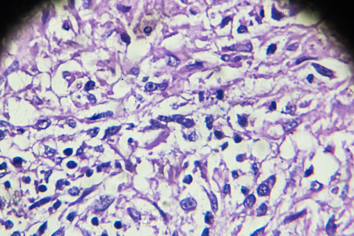
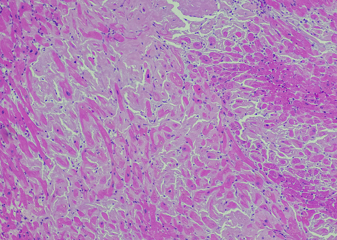
Free Images: "bestof:Chlamydia pneumoniae.jpg en Chlamydia pneumoniae in epithelial cell Acute bronchitis 1 - infected epitheliocyte 2 - uninfected epitheliocytes 3 - chlamydial"
Terms of Use   Search of the Day