Click Here for More Images from iStock- 15% off with coupon 15FREEIMAGES 
Nodularia sp. toxic algae under microscopic view, nitrogen-fixing cyanobacteria, cyanotoxin, harmful to humans, x40
plant Pine Cone-Female L.S. under light mircoscope with white background
Microscopic photo of a thin section of calcareous tufa of Holocene age.
Photomicrograph of filamentous blue-green algae, Oscillatoria species. Each band along the stand is one cell. This filament is very short probably having recently parted from a longer one. Filaments are typically many times longer. This algae is mobile, it moves and twists through the water. San Francisco Bay, California, USA. Live specimen. Wet mount, 40X objective, transmitted brightfield illumination. Note - motion blur of live specimen, very shallow depth of field, chromatic aberration and uneven focus are inherent in light microscopy.
Epidermis. Electron microscope micrograph showing a keratinocyte of spinous layer. The epithelial cell has a polygonal shape, central nucleus with nucleolus, cytoplasm full of keratin filament bundles, and numerous dark desmosomes crossing the intercellular spaces.
Culture of Clostridium difficile, an anaerobic bacterium. This bacterium is in some cases able to cause severe diarrhoea leading to pseudomembranous colitis and death. Clostridium difficile has caused many nosocomial outbreaks in hospitals.
centropyxis aculeata (amoeboid organism) under the microscope - optical microscope x400 magnification
Cheyletiella blakei Mite - Small Cat pest view under a microscope
Licmophora sp. algae, marine and freshwater diatom under microscopic view. Genus of benthic, photosynthetic and epiphyte diatom
Urothelial carcinoma, high grade.  High-grade tumors have an aggressive appearance under a microscope and are assumed invasive in the kidney or ureter.
Cross section of Escherichia coli bacteria
Particles, medical concept
Paramecium is a genus of unicellular ciliated protozoa,  Paramecia are widespread in freshwater, brackish, and marine environments and are often very abundant in stagnant basins and ponds.
plant PomeTomato Flesh W.M. under light mircoscope with white background
Planarian parasite (flatworm) under microscope view.
Different germs in the human intestines called microbiome,Bacteria Lactobacillus in human intestine,Beneficial healthy intestinal bacterium microflora,Gut bacteria
Transmission electron micrograph (TEM) showing a striated skeletal muscle cell in cross section. Among myofibrils (sectioned at the band A level), mitochondria and sarcoplasmic reticulum can be seen.
Organic abstract image of a frost tipped hydrangea flower that has been heavily post processed in Lightroom and Topaz studios.
Microalgae under microscopic view, green algae, cyanobacteria, phytoplankton, diatom, algae mix collage background
Cheyletiella blakei Mite - Small Cat pest view under a microscope
E. coli stands for Escherichia coli is a bacteria that is commonly found in the lower intestine of warm-blooded organisms 3d rendering
Escherichia Coli (E. Coli.)
Host cells with spores (mold) are inside wood under the microscope for education.
Blastomycosis is a fungal infection caused by inhaling Blastomyces dermatitidis spores. If it involves only the lungs, it is called pulmonary blastomycosis. Only about half of people with the disease have symptoms, which can include fever, cough, night sweats, muscle pains, weight loss, chest pain, and feeling tired.
Moss leaf under the microscope, green cell background
Liver Fluke Section under light microscope with white background
Transmition Electron microscopy of an epithelial cell where mitochondria of several sizes can be seen with the characteristic double membrane and internal cristaes. Mitochondria are believed to be of bacteria origin. It has its own genome and RNA and protein which are similar to those of bacteria.
Natural agate surfaces, suitable for backgrounds and wallpapers.
Microscopic of paramecium and amoeba
West Nile are invading blood system in blood vessels. Easy to crop for all your social media and print sizes.
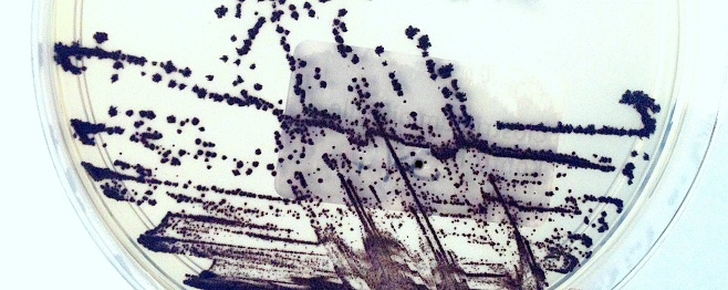
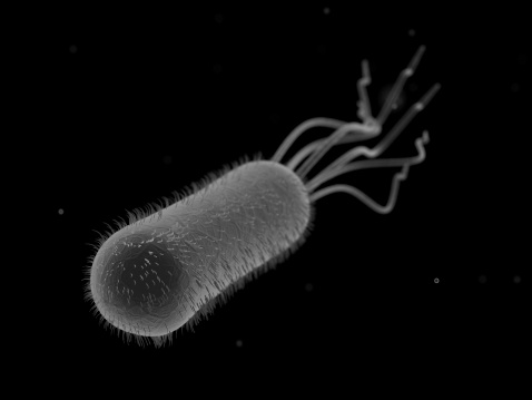
Free Images: "bestof:Campylobacter jejuni 5778 lores.jpg This scanning electron micrograph depicts a number of Gram-negative Campylobacter jejuni bacteria magnified 11 734x C jejuni"
hela-cells-bacteria-germs-virus-544318.jpg
Campylobacter_jejuni_5778_lores.jpg
MRSA_SEM_7821_lores.jpg
Multidrug-resistant Klebsiella pneumoniaeand neutrophil.bmp.jpg
Scanning_Electron_Micrograph_of_a_Flea.jpg
Ralstonia_mannitolilytica.png
Yersinia_pestis_Bacteria.jpg
Nanoknife.jpg
Blood clot in scanning electron microscopy.jpg
Clostridium_difficile_EM.png
Malassezia_furfur_SEM_lores.jpg
Nanoparticles; scanning electron micrograph (5887833046).jpg
Streptococcus_pyogenese_Bacteria.jpg
Yersinia_pestis.jpg
Giardia_1.jpg
Spermatozoa-human-3140x.jpg
Spermatozoa-human-1000x.jpg
Anthrax spores (5940425745).jpg
Nipah.jpg
Streptococcus_pneumoniae-263.jpg
White_blood_cell_with_MRSA.jpg
Bosch process sidewall.jpg
Micrococcus_luteus_9758.jpeg
Electron_Micrograph_of_Flea_derivate.jpg
Malaria_Plasmodium_gallinaceum.jpg
TEM_phil_CDC_11635_lores.jpg
Bone Cells (5828753702).jpg
Malaria-infected_Red_Blood_Cell.jpg
Tin Spheres (5828754970).jpg
Beryllium Reference Material (5940427681).jpg
TEM of L-form bacteria-Mark Leaver Newcastle University.tif
TEM of L-form bacteria-Mark Leaver Newcastle University.jpeg
Wide field EM of L-form bacteria-Mark Leaver Newcastle University.jpeg
Wide field EM of L-form bacteria-Mark Leaver Newcastle University.tif
Schistosome_Parasite_SEM.jpg
New Quantum Dot Transistor Counts Individual Photons (5941062028).jpg
Scanning Electron Microscope with Spin Polarization Analysis (5941086294).jpg
Terms of Use   Search of the Day