Click Here for More Images from iStock- 15% off with coupon 15FREEIMAGES 
3D rendering of human cerebrum left and right hemispheres x-ray style shading, glowing star field within head and skull, top view.
A meningioma is a tumor that arises from the meninges — the membranes that surround the brain and spinal cord. Although not technically a brain tumor, it is included in this category because it may compress or squeeze the adjacent brain, nerves and vessels.
Dopamine is a chemical messenger and neurotransmitter produced in the brain that affects many bodily functions. It's an organic chemical that's part of the catecholamine and phenethylamine families. Dopamine is responsible for many effects, including mood, movement, memory, motivation, attention.
Human brain MRI.
Gut-brain connection or gut brain axis. Concept art showing a connection from the gut to the brain. 3d illustration.
Amygdalas in a human brain, part of the limbic system,
The corpus callosum , also callosal commissure, is a wide, thick nerve tract, consisting of a flat bundle of commissural fibers, beneath the cerebral cortex in the brain. The corpus callosum is only found in placental mammals.It spans part of the longitudinal fissure, connecting the left and right cerebral hemispheres, enabling communication between them.
Magnetic resonance images of the brain (MRI brain). Computed resonance imaging of the human brain.
Human brain nervous system anatomy, medical diagram with parasympathetic and sympathetic nerves. medically accurate, skull cross section, Sebaceous bulb, 3d render
WHO grade I menWorld Health Organization (WHO) grade I meningiomas are intracranial extracerebral tumors, in which microsurgery as a stand-alone therapy provides high rates of disease control and low recurrence rates. ingioma.
3D Rendering of male pelvis, hip, leg bones and ligaments labeled on a white background.  Front view.
Human brain MRI and AI technology.
3d rendering medical illustration of a female Brain anatomy PITUITARY GLAND - cross section
Medical illustration about Sacrum.3d rendering. No AI create.
The nervous system is a highly complex part of an animal that coordinates its actions and sensory information by transmitting signals to and from different parts of its body. The nervous system detects environmental changes that impact the body, then works in tandem with the endocrine system to respond to such events.
X-ray image of a human brain made up mostly of water
Human brain anatomy, basal ganglia. 3D illustration showing caudate nucleus (green), putamen (yellow), and lateral ventricles (blue)
Epithelioid mesothelioma is a cancer subtype caused by asbestos. It is the most common mesothelioma cell type, accounting for 50% to 70% of cases.
The sacrotuberous ligament (STL) is a stabiliser of the sacroiliac joint and connects the bony pelvis to the vertebral column 3d illustration
The central nervous system is made up of the brain and spinal cord 3D illustration
Digital medical illustration: Posterior (back) view (orthogonal) of human skull (includes teeth) and neck featuring vertebrae, discs and ligaments (includes visible Interspinale, Nuchae, Anulus, Flavum, Capsula and Intertransversarium).
isolated brain image . Human brain model from 3 different angles
Web of connections grows inside a brain.
3D Illustration Concept of
Serous carcinoma is the most common type of ovarian cancer, accounting for approximately 75% of epithelial ovarian cancers.
The human brain has many properties that are common to all vertebrate brains, including a basic division into three parts called the forebrain, midbrain, and hindbrain, each with fluid-filled ventricles at their core, and a set of generic vertebrate brain structures including the medulla oblongata, pons, cerebellum, optic tectum, thalamus, hypothalamus, basal ganglia, olfactory bulb, and many others.
Section of epididymis under light microscope with white background
Computer Generated Image: Sideview of a transparent head of a man with the brain parts isolated and labeled against a white background.
Human brain MRI and AI technology.
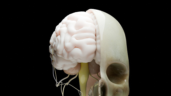
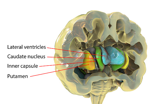
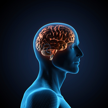
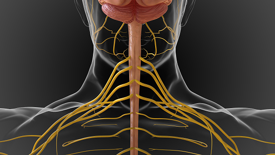
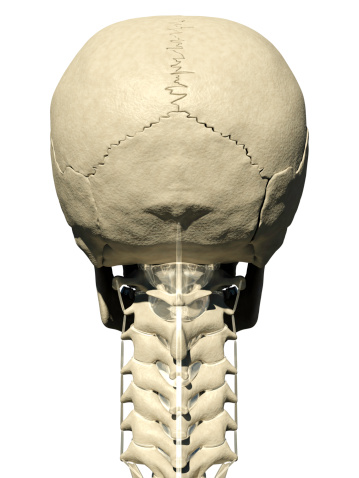
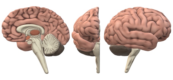
Free Images: "bestof:Brain and Nearby Structures.png en The brain and nearby structures including the skull meninges ventricles and spinal cord An enlarged inset shows the skull"
oakland-skyscraper-mirroring-49549.jpg
skull-brain-head-science-medical-1209733.jpg
anatomy-woman-human-face-body-254120.jpg
Decomposing cow skull.jpg
anatomy-man-human-face-body-lips-254129.jpg
woman-human-pair-man-face-body-254133.jpg
skeleton-bones-human-body-anatomy-1480319.jpg
A boy poses with a pelican skull.jpg
Facilities and structures after windy storm.jpg
Buildings facilities and structures after hurricane.jpg
Metal structures demaged after hurricane.jpg
appearance-bleak-body-bones-brain-216187.jpg
anatomy-background-body-bone-brain-71734.jpg
Employee talks with visitors about the pelican skull.jpg
Dwarf crested iris flower in field plant.jpg
Girl holding the skull of a pelican.jpg
Facilities and structures in field left out after hurricane.jpg
anatomy-background-body-bone-brain-71732.jpg
Pachyprosopis_cornuta,_f,_australia,_back_2014-11-02-01.15.17_ZS_PMax.jpg
Tubal Pregnancy with embryo.jpg
2016_Bitner_Ranch_Root_Cellar_Project.jpg
Pyramid in long beach.jpg
Overgrown barn.jpg
Pyramids ridge near Nanutarra.jpg
Wooden fence on sand dunes area.jpg
Wooden dorey boat in an Alaskan meadow.jpg
me_myself_and_i....jpg
Historic blacksmith shop and railroad tie house.jpg
Reflections in lake in marsh.jpg
Reflection of a bird box.jpg
Gray769-en.svg
Gray769-es.svg
Gray769-pt.svg
Complete neuron cell diagram numbered large numbers.svg
Basal_Ganglia_and_Related_Structures_ru.svg
Basal_Ganglia_and_Related_Structures_no.svg
Basal_Ganglia_and_Related_Structures_es.svg
Basal_Ganglia_and_Related_Structures.svg
BrainCaudatePutamen.svg
Complete neuron cell diagram pl.svg
Complete neuron cell diagram es.svg
Complete neuron cell diagram it.svg
Complete neuron cell diagram without text.svg
Complete neuron cell diagram en.svg
Complete neuron cell diagram ru.svg
Central nervous system.svg
End_of_Knowles'_action_off_Havana,_1_October_1748.jpg
Dr_Deijman’s_Anatomy_Lesson_(fragment),_by_Rembrandt.jpg
Stem cell treatments.svg
brain-stem-cerebrum-spinal-cord-305774.svg
brain-head-skull-cranium-1197343.svg
brain-head-skull-cranium-1197344.svg
skull-cogs-thinking-human-brain-1529065.png
matte-painting-skull-sea-brain-1685255.jpg
computer-tomography-ct-radiography-62942.jpg
mri-magnetic-x-ray-skull-head-782457.jpg
mri-magnetic-x-ray-skull-head-782459.jpg
drawing-nerve-cell-neurone-732830.png
drawing-nerve-cell-neurone-730778.jpg
drawing-nerve-cell-neurone-730755.png
brain-cranium-head-psychology-1299830.svg
halloween-anatomy-background-body-193532.jpg
halloween-anatomy-background-body-193531.jpg
skull-diagram-labelled-human-41552.svg
skull-anatomy-head-human-health-1132246.png
skull-diagram-head-human-medical-40843.svg
Homo erectus.svg
Homo ergaster.svg
Homo habilis.svg
Australopiteco.svg
Nearby Stars (14ly Radius) ltz.svg
colorful-prismatic-chromatic-1325265.svg
colorful-prismatic-chromatic-1325268.svg
Homo antecessor.svg
head-brain-radiology-medical-human-1058432.jpg
Complete_St_Wenceslas_skull.jpg
6 week human embryo nervous system.svg
Complete neuron cell diagram zh.svg
Complete neuron cell diagram pt.svg
Complete neuron cell diagram numbered.svg
Complete neuron cell diagram zh-hant.svg
Basal_Ganglia_lateral.svg
Rattus_macleari_skull_labeled.svg
Basal_Ganglia_lateral-el.svg
Brain_areas_implicated_in_Tourette_syndrom.svg
Gray770-ar.svg
Buildings facilities and structures old photograph.jpg
Epidural blood patch.svg
Gray770-en.svg
Malloc versus HeapAlloc.svg
Girl_and_Skull.jpg
Brain_areas_implicated_in_Tourette_syndrom-el.svg
skull-heart-death-soul-mind-1391722.jpg
skeleton-skull-death-eternity-1243818.jpg
Dsa k way merge problem lists.svg
Canal Fall.svg
Gray770-pt.svg
GJ581orbits Vogt2010.svg
Gray770-es.svg
La_Iglesia_de_Nuestra_Señora_Reina_de_los_Angeles.jpg
Terms of Use   Search of the Day