Click Here for More Images from iStock- 15% off with coupon 15FREEIMAGES 
Chest CT SCAN of female patient with breast protesis and lung infection
Oviduct T.S. under light microscope with white background
normal head  scan brain window
Magnetic resonance images of the brain (MRI brain). Computed resonance imaging of the human brain.
brainstem is the bottom, stalklike portion of your brain.
CAT scan of human abdomen...man with aneurysm of abdominal aorta
Enlarged Prostate organ gland as Benign Prostatic Hyperplasia as a urology medical illustration concept for part of the male reproductive anatomy and lower urinary tract symptoms.
A Computed Axial Tomography scan of my brain.  Taken with contrast.  Great image for medical applications.
isolated brain image . Human brain model from 3 different angles
Metastatic neuroendocrine tumor. Site: Liver.  Neuroendocrine tumors are cancers that begin in specialized cells called neuroendocrine cells. Neuroendocrine cells have traits similar to those of nerve cells and hormone-producing cells. Neuroendocrine tumors are rare and can occur anywhere in the body.
Medical Imaging and Radiology
Human brain MRI.
Prostate cancer, 3D illustration showing tumor inside prostate gland and closeup view of cancer cells
Pine Root C.S. under light mircoscope with white background
CAT Scan of young female with breast prosthesis and left upper lobe pulmonary tuberculosis. Medical themes
Magnetic resonance images of the brain (MRI brain). Computed resonance imaging of the human brain.
forming of underskin cellulite illustration
Serous carcinoma is the most common type of ovarian cancer, accounting for approximately 75% of epithelial ovarian cancers.
Adipose tissue human, Smooth muscles human, Bone human and Striated (skeletal) muscle human under the microscope in Lab.
The diaphragm is a muscle that helps you inhale and exhale (breathe in and out) 3d illustration
Mid-sagittal section showing the anatomy of the pelvis and testis. The model shot in studio environment. Isolated on white background.
CT scan of Chest axial view for diagnostic Pulmonary embolism (PE) , lung cancer and covid-19.
CT of thorax of four patients having covid-19 disease. Four variations are seen on four lung x-ray images. Shot with a full frame mirrorless camera.
Cat Scan of female patient with centrilobular emphysema. Medical themes
Squamous cell carcinoma of the lung is a type of lung cancer. It occurs when abnormal lung cells multiply out of control and form a tumor.
Human brain anatomy, basal ganglia. 3D illustration showing caudate nucleus (green), putamen (yellow), and lateral ventricles (blue)
Human brain MRI and AI technology.
CT Scan of an abdomen of a patient with abdominal pain showing swollen appendix suggesting the diagnosis of acute appendicitis.
Digital medical illustration: Posterior (back) view (orthogonal) of human skull (includes teeth) and neck featuring vertebrae, discs and ligaments (includes visible Interspinale, Nuchae, Anulus, Flavum, Capsula and Intertransversarium).
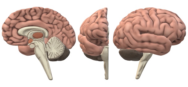
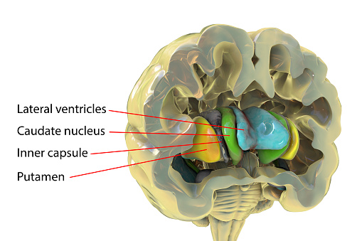
Free Images: "bestof:Basal Ganglia and Related Structures-IT.png Author - John Henkel from the Food and Drug Administration Structure of the basal gandlia including thalamus globus"
Basal_Ganglia_and_Related_Structures.svg
Basal_Ganglia_and_Related_Structures_ru.svg
Basal_Ganglia_and_Related_Structures_no.svg
Basal_Ganglia_and_Related_Structures_es.svg
BrainCaudatePutamen.svg
Brain_areas_implicated_in_Tourette_syndrom.svg
Brain_areas_implicated_in_Tourette_syndrom-el.svg
Basal_Ganglia_lateral.svg
Basal_Ganglia_lateral-el.svg
Basalganglia.svg
Whitehall_PORTICO_OVERLOOKING_THE_BAY.jpg
Whitehall_LAND_SIDE.jpg
Receptacle growths gynophore androgynophore anthophore androphore hypanthium compare with stipitate gynoecium and monadelphous stamens definitions as in APWeb.svg
Cannelton_Cotton_Mill_065589pr.jpg
Basal_Ganglia_and_Related_Structures-IT.png
Brain_structure.png
Basal-ganglia-2.jpg
Pallidus.jpg
Telencephalon-Horiconatal.jpg
Anatomie-Basalganglien-A.jpg
Internal view of basal ganglia.jpg
External view of basal ganglia.jpg
Putamen.jpg
Caudate nucleus.jpg
Basalganglier.png
Basalganglien.png
Anterior face of brainstem.jpg
Posterior face of brainstem.jpg
External face of brainstem.jpg
Parkinsons, huntingtons.png
Basale ganglia circuits.png
Gray741.png
Center_for_Veterinary_Medicine_logo.png
Cosmetics Claims.jpg
BGA.PNG
Campaign Against Rats (FDA 188).jpg
Pseudohypoparathyroidism.gif
John_E_Sheridan_WWI_US_Food_is_Ammunition_Dont_Waste_it.jpg
FDA History - Dr. Kelsey and John F. Kennedy.jpg
President_Signing_the_1962_Drug_Amendments.jpg
Inspecting_eggs.jpg
Guiding Public Health Professionals to Online Resources.jpg
John_F._Kennedy_Telegram_to_Nat_King_Cole_March_11,_1960_-_NARA_-_194003.jpg
John_F._Kennedy_Telegram_to_Nat_King_Cole_March_11,_1960_-_NARA_-_194003.tif
Colima_-_Group_of_Figurines_and_Architectural_Model_-_Walters_20092033_-_Three_Quarter.jpg
Colima_-_Group_of_Figurines_and_Architectural_Model_-_Walters_20092033_-_Three_Quarter_Back.jpg
Colima_-_Group_of_Figurines_and_Architectural_Model_-_Walters_20092033.jpg
Colima_-_Group_of_Figurines_and_Architectural_Model_-_Walters_20092033_-_Left_Side.jpg
Colima_-_Group_of_Figurines_and_Architectural_Model_-_Walters_20092033_-_Group.jpg
Colima_-_Group_of_Figurines_and_Architectural_Model_-_Walters_20092033_-_Three_Quarter_Back_(2).jpg
Colima_-_Group_of_Figurines_and_Architectural_Model_-_Walters_20092033_-_Three_Quarter_Front.jpg
Colima_-_Group_of_Figurines_and_Architectural_Model_-_Walters_20092033_-_Right_Side.jpg
Colima_-_Group_of_Figurines_and_Architectural_Model_-_Walters_20092033_-_Three_Quarter_Front_(2).jpg
Colima_-_Group_of_Figurines_and_Architectural_Model_-_Walters_20092033_-_Back.jpg
Colima_-_Group_of_Figurines_and_Architectural_Model_-_Walters_20092033_(2).jpg
Colima_-_Group_of_Figurines_and_Architectural_Model_-_Walters_20092033_-_Three_Quarter_(2).jpg
Colima_-_Group_of_Figurines_and_Architectural_Model_-_Walters_20092033_-_Back_(2).jpg
Colima_-_Group_of_Figurines_and_Architectural_Model_-_Walters_20092033_(2)_(3).jpg
Colima_-_Group_of_Figurines_and_Architectural_Model_-_Walters_20092033_-_Three_Quarter_(2)_(3).jpg
Colima_-_Group_of_Figurines_and_Architectural_Model_-_Walters_20092033_-_Side_A.jpg
Colima_-_Group_of_Figurines_and_Architectural_Model_-_Walters_20092033_-_Back_(2)_(3).jpg
Colima_-_Group_of_Figurines_and_Architectural_Model_-_Walters_20092033_-_Three_Quarter_Back_(2)_(3).jpg
Colima_-_Group_of_Figurines_and_Architectural_Model_-_Walters_20092033_-_Three_Quarter_Front_(2)_(3).jpg
Colima_-_Group_of_Figurines_and_Architectural_Model_-_Walters_20092033_-_Three_Quarter_Left.jpg
Colima_-_Group_of_Figurines_and_Architectural_Model_-_Walters_20092033_-_Three_Quarter_Right.jpg
Colima_-_Group_of_Figurines_and_Architectural_Model_-_Walters_20092033_-_Left_Side_(2).jpg
The_Quarterly_journal_of_the_Geological_Society_of_London_(13960146853).jpg
The_Quarterly_journal_of_the_Geological_Society_of_London_(12893785694).jpg
British_Entomology_Volume_7_(John_Curtis)_Plate_417.jpg
Treblinka II aerial photo (1944).jpg
Bud_Uanna_A_TEAM_INTELLIGENTSIA_Toward_a_"Thought_Police_State"_p.1.jpg
M2-F1_lifting_body_and_Paresev_1B_on_ramp_DVIDS735192.jpg
Terms of Use   Search of the Day