Click Here for More Images from iStock- 15% off with coupon 15FREEIMAGES 
Transmission electron microscope (TEM) micrograph showing a brush border in longitudinal section (inset: light microscope appearance). The microvilli are parallel and tightly packed.
Diffuse large B-cell lymphoma, or DLBCL, is a cancer that starts in white blood cells called lymphocytes.
Backgrounds of Bacteria and Mold under the microscope in Lab.
Yeasts sprout in microscopy
A micrograph of nanoparticles made of lipids containing an anticancer drug. The drug appears as a crystal inside of the spherical liposomes
Photomicrograph of rotifer. Rapidly rotating cilia at top of head, internal organs visible. Live specimen. Wet mount, 40X objective, transmitted brightfield illumination.
Particles, medical concept
Leiden leaf c.s. under microscope
Epidermis. Electron microscope micrograph showing a keratinocyte of spinous layer. The epithelial cell has a polygonal shape, central nucleus with nucleolus, cytoplasm full of keratin filament bundles, and numerous dark desmosomes crossing the intercellular spaces.
Metastatic prostatic adenocarcinoma
Transmition Electron microscopy of an epithelial cell where mitochondria of several sizes can be seen with the characteristic double membrane and internal cristaes. Mitochondria are believed to be of bacteria origin. It has its own genome and RNA and protein which are similar to those of bacteria.
plant Flower bud of Lilium brownii var.viridulum T.S.\nunder light mircoscope with white background
Histopathology of diffuse sclerosing glomerulonephritis, light micrograph, photo under microscope
Slice of natural crystal for patterns and textures use with high resolution.
Symbolic illustration of infectious viruses under the microscope, for example influenza viruses, smallpox, measles or similar. Medical research and investigation of diseases and healthcare concept.
Brush Grunge Background
Transmission electron micrograph (TEM) showing a striated skeletal muscle cell in cross section. Among myofibrils (sectioned at the band A level), mitochondria and sarcoplasmic reticulum can be seen.
Burkitt lymphoma is a form of non-Hodgkin's lymphoma in which cancer starts in immune cells called B-cells. Recognized as a fast growing human tumor, Burkitt lymphoma is associated with impaired immunity and is rapidly fatal if left untreated.
Abstract gray sharpening wet stone background. Gray abstract texture. Blank grunge dark gray background for text.
Digital Dispersed corona virus on petri dish. Animation of multiplication of CORONA VIRUS COVID-19
PAPS Smear study of a young women under microscopy showing Atrophic changes in uterus which is unusual at young age.
Photomicrograph of two rotifers. Rapidly rotating cilia at top of heads, internal organs visible. Live specimen. Wet mount, 10X objective, transmitted brightfield illumination.
An electron image of several nanoparticles made of lipids (liposomes) containing an anticancer drug (doxorubicin)
Abs Bubonic plague bacteria - 3d rendered image structure view on black background. \nPlague Infection concept.  Hologram bacteria view.
Ink splatter. Blur ornament. Defocused black white paint splash dirt stains texture graphic pattern grunge abstract art illustration background.
Liver Fluke Section under light microscope with white background
Transmission electron microscope (TEM) micrograph showing dark melanin granules in a melanocyte (top center) and in several keratinocytes, which, unlike melanocytes, have bundles of keratin filaments
Thymoma thymus.  Thymoma and thymic carcinoma are diseases in which malignant (cancer) cells form in the thymus. Thymoma is linked with myasthenia gravis and other autoimmune paraneoplastic diseases.
Light photomicrograph of pine tree wood seen through a microscope
Luxury stone background, nature pattern for use.
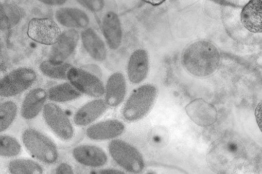
Free Images: "bestof:B00528-Swine-flu.png en This negative stained transmission electron micrograph TEM depicted some of the ultrastructural morphology of the A/CA/4/09 swine flu"
B00528-Swine-flu.png
H5N1_virions_108,000x.jpg
A_(H5N1)_virion,_a_type_of_bird_flu_virus_which_is_a_subtype_of_avian_influenza_A.jpg
Nipah.jpg
Chikungunya_virus_particles-PHIL-17369.jpg
TEM_phil_CDC_11635_lores.jpg
Micrococcus_luteus_9758.jpeg
TEM of L-form bacteria-Mark Leaver Newcastle University.tif
TEM of L-form bacteria-Mark Leaver Newcastle University.jpeg
Coronavirus.jpg
MERS_coronavirus.jpg
Eosinophil TEM.jpg
Rotavirus_TEM_B82-0337_lores.jpg
H1N1_virus_particles.jpg
TEM of silicon nanowire (5887830328).jpg
Chromatin nucleofilaments.png
Diseases of Swine 23-3.png
H1N1_virus_particles_II.jpg
Staph_aureus_outside_a_white_blood_cell.jpg
Transmission Electron Microscope Kemira.jpg
Neodymium Crystal Structure Nd2Fe14B.jpg
Deinococcus_radiodurans.jpg
MRSA_SEM_7821_lores.jpg
Poxvirus_of_molluscum_contagiosum_TEM_B81-166_lores.jpg
Wide field EM of L-form bacteria-Mark Leaver Newcastle University.tif
Giardia lamblia.bmp.jpg
Wide field EM of L-form bacteria-Mark Leaver Newcastle University.jpeg
25K15pA9Def4sec Arman 4 Box1.png
Vibrio cholerae.jpg
Blood clot in scanning electron microscopy.jpg
Countries with confirm double cases of swine flu.png
Retino ME con sezioni.jpg
Multidrug-resistant Klebsiella pneumoniaeand neutrophil.bmp.jpg
Cilia.jpg
Bacillus subtilis.jpg
Kamiakin.jpg
Nez.Perce.Camp.Lapwai.1899.jpg
Mission.Spalding.jpg
Chief.Joseph.Band.1877.jpg
Transmission line animation open short.gif
Terms of Use   Search of the Day