Click Here for More Images from iStock- 15% off with coupon 15FREEIMAGES 
3d render of T cells attacking cancer cells
Digital background depicting innovative technologies in medicine systems, neural interfaces and research genetic and biology
The Raf/MEK/ERK (MAPK) propagates growth signals in the cell. Misregulation can lead to cancer.\nThe red dots on the active EGFR are phosphorylated tyrosines. PDB entries: 1egf, 1nql, 1m17, 2jwa, 3njp, 2gs6, 1gri, 1xd2, 3ksy, 5p21, 6xi7, 6q0j, 2y4i,
Bacterial cell or virus, 3d generated view from microscope.
Follicular lymphoma is the most common type of of low-grade non-Hodgkin lymphoma (NHL). It develops when white blood cells cluster together to form lumps in your lymph glands or organs. There are two main types of lymphoma: Hodgkin lymphoma and non-Hodgkin lymphoma.
Haematocysts of single-celled green algae,
Cells in the body examined by endoscope
B-chain pentamer and A-chain subunit (green), 3D cartoon and Gaussian surface model, white background
prostate cancer, prostatic hyperplasia, Structure of male prostate, cross section, 3d illustration
3D rendering of microscopic molecular structure
Bacterial dehalogenases remove halogen atoms from organic compounds. The one shown here is a reductive dehalogenase from PDB entry 4ur0. It uses a cobamide cofactor (orange) and iron-sulfur clusters to reduce the substrate. Dehalogenases may be used as a way to clean up areas polluted with halogenated compounds.
H2 Hydrogen Molecule Fuel Cell Element Vehicle Biodegradable Ethical Renewable Energy
Coronavirus, cancer cell or embryonic stem cell. Medical research or pandemic prevention banner with microscopic disease image. Red bacteria or virus in clear mucus on blue background. 3d illustration
Stem of Nerium indicum T.S. under light mircoscope with white background
Green algae cells background
Endosomes - 3d rendered image of Endosomes are membrane-bound organelles. They are parts of endocytic membrane transport pathway.  Endomembrane system in cells. Bilayer Lipid Membrane. Abstract medical illustration. Microscopic view. Microbiologic research concept.
3d render of cerebral organoids
stx2a is a subtype of Shiga toxin-producing E. coli (STEC) linked to Hemolytic Uremic Syndrome (HUS), a severe condition impacting kidney function and blood clotting. STEC poses a significant risk of foodborne illness and can spread within communities including family members.
The cellular engineering is mainly associated with the modifications and alterations of host cells for enhancing the yield and quality of the expressed protein 3d rendering
Digital background depicting innovative technologies in medicine systems, neural interfaces and research genetic and biology
Nuclear fusion reaction generating energy and light. Digital illustration.
Photomicrograph of two rotifers. Rapidly rotating cilia at top of heads, internal organs visible. Live specimen. Wet mount, 10X objective, transmitted brightfield illumination.
3D cartoon and Gaussian surface models, chain id color scheme, PDB 6jod, putative membrane is shown, white background
Inside protein molecule
Endosomes - 3d rendered image of Endosomes are membrane-bound organelles. They are parts of endocytic membrane transport pathway.  Endomembrane system in cells. Bilayer Lipid Membrane. Abstract medical illustration. Microscopic view. Microbiologic research concept.
Cytochrome P450 molecule. CYP 3A4 is the most promiscuous of the human CYP enzymes. Molecular model. 3D rendering. Illustration
Main mutation site of the coronavirus variant B.1.531 emerged in South Africa. The spike protein (red) is bound to the angiotensin converting enzyme 2 (blue). The mutation is at the amino acid position 484 (yellow). Source: PDB entry 6cs2.
Covid-19 Coronavirus Variants Omicron Delta Gamma Beta Alpha Pandemic Virus Disease Mutation
Cysts of Entamoeba coli protozoan, 3D illustration. E. coli is a non-pathogenic ameba, its cyst is 15-25 mkm, has 8 nuclei and chromatoidal bar elongated with splintered ends (shown red)
Urothelial carcinoma, high grade.  High-grade tumors have an aggressive appearance under a microscope and are assumed invasive in the kidney or ureter.
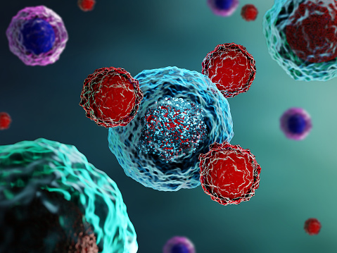
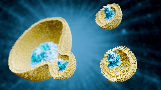
Free Images: "bestof:B cell activation.svg en When a B cell encounters its triggering antigen it gives rise to many large cells known as plasma cells Every plasma cell is"
Euryglossidia,_f,_australia,_face_2014-10-11-17.16.19_ZS_PMax.jpg
Euryglossidia,_f,_australia,_side_2014-10-11-17.23.14_ZS_PMax.jpg
Humanstemcell.JPG
dasypoda,f,_united_kingdom,_side_2014-10-11-20.33.26_ZS_PMax.jpg
dasypoda,f,_united_kingdom,_face_2014-10-11-20.28.43_ZS_PMax.jpg
dasypoda,f,_united_kingdom,_back_2014-10-11-20.21.29_ZS_PMax.jpg
bee_19992,_f,_face,_kenya_2014-08-07-14.58.57_ZS_PMax.jpg
bee_19992,_f,_angle,_kenya_2014-08-07-14.44.23_ZS_PMax.jpg
Catopsilia pomona 3 by kadavoor.jpg
Large cell tower.jpg
paprika-fruit-bowl-chromoplasts-1582256.jpg
microscope-sea-urchin-egg-splitting-1276131.jpg
Project_365_#287:_141009_A_Battery_of_Batteries.jpg
jail-prison-police-crime-law-429633.jpg
jail-prisoner-captive-police-crime-429639.jpg
jail-cells-jail-penitentiary-police-429638.jpg
Tissue Engineering; Biomaterials for the Heart (5881107872).jpg
agar-breeding-ground-red-60571.jpg
8-cell-simple.gif
cellular_repeater.jpg
gliding-glider-glider-pilot-1422785.jpg
gliding-glider-glider-pilot-1422784.jpg
Tree_simulation.jpg
Hotel_à_abeilles_solitaires_-_Bees_motel.jpg
Seafood fishing boat greece.jpg
The_Importance_of_Platinum_Elements.jpg
Solar garden light.jpg
Secretary_Kerry_Poses_for_a_Photo_with_a_Baby_Elephant_at_the_Sheldrick_Elephant_Orphanage_(17358471511).jpg
Colletes_phaceliae,_f,_back,_Pennington_Co,_South_Dakota_2015-12-01-12.06.jpg
Colletes_phaceliae,_f,_side,_Pennington_Co,_South_Dakota_2015-12-01-12.40.jpg
Colletes_phaceliae,_f,_face,_Pennington_Co,_South_Dakota_2015-12-01-12.23.jpg
Camaecyparis_lawsoniana.jpg
SLNSW 13194 Women volunteer workers doing up clothing parcels at the ACF rooms taken for Australian Comforts Fund.jpg
B_cell_activation.svg
B_cell_activation_-_ru.svg
T_cell_activation_-_ru.svg
T_cell_activation.svg
Desmosome cell junction en.svg
portugues-sexo_alemão-sex_ingles-sex_africano-seks_russo-секс_albanes-sex_chines-性別_bulgaro-секс_croata-spol_gales-rhyw_coreano-섹스_frances-sexe_japones-セックス_filandes-sukupuolesta_arabe-الجنس_prontos_ai_tens_em_15_linguas_a_p.jpg
Cell membrane detailed diagram en.svg
Cell membrane detailed diagram blank.svg
Endocytosis types-fr.svg
Complete neuron cell diagram numbered large numbers.svg
Complete neuron cell diagram en.svg
Eukaryotic cilium diagram en.svg
Complete neuron cell diagram it.svg
Cellular tight junction keys.svg
Cell membrane detailed diagram id.svg
Insulin glucose metabolism ZP.svg
Insulin glucose metabolism ZP-ca.svg
cells-cell-structure-organism-blood-75305.jpg
Antigen_presentation_alt.svg
Animal mitochondrion diagram unlabelled.svg
Animal mitochondrion diagram en (edit).svg
Endocytosis types.svg
Desmosome cell junction it.svg
Complete neuron cell diagram es.svg
Complete neuron cell diagram without text.svg
Complete neuron cell diagram pl.svg
Endocytoza typy.svg
Complete neuron cell diagram ru.svg
Thyroid hormone synthesis.svg
NK-cell activation.svg
Design_Analysis_of_the_B-24J_Liberator.png
Average prokaryote cell- en.svg
Animal cell structure en.svg
24-cell duals.svg
Dendritic Cell ZP.svg
Scheme simple diffusion in cell membrane-en.svg
Stem cell treatments.svg
Animal Cell Unannotated.svg
1829 Braille cell numbering.svg
Taste bud cell types.svg
Titaniumdioxide unit cell rutile.svg
Photovoltaics cell production.svg
Oncogene activation illustration.jpg
F4 roots by 24-cell duals.svg
UnitCell.svg
Outer hair cell and Deiter's cell.svg
Complete neuron cell diagram numbered.svg
Ebola Pathenogensis.svg
Scheme simple diffusion in cell membrane-es.svg
Scheme simple diffusion in cell membrane-de.svg
Tipica cellula procariote.svg
Scheme simple diffusion in cell membrane-fr.svg
Solid oxide fuel cell protonic.svg
Prokaryote cell diagram international.svg
Animal cell structure numbered version.svg
Complete neuron cell diagram pt.svg
Ebola Pathenogensis path.svg
Complete neuron cell diagram zh.svg
Animal cell structure uk.svg
Complete neuron cell diagram zh-hant.svg
Animal cell structure ca.svg
Endosymbiosis.svg
Ebola Pathenogensis path-hu.svg
Plasmolyse Pflanzenzelle.svg
Zeppelin-LZ-127 gas cells.svg
Terms of Use   Search of the Day