Click Here for More Images from iStock- 15% off with coupon 15FREEIMAGES 
A dysgerminoma is a tumor of the ovary that is composed of primitive, undifferentiated germ cells. Germ cell tumors arise from primordial germ cells of the ovary ; however, pathogenesis of the ovarian germ cell tumors is unknown.
forming of underskin cellulite illustration
The vas deferens is part of the male reproductive system, along with the scrotum .
corn stem micrograph with dye
The study of plant tissues under the microscope in the laboratory.
Mid-sagittal section showing the anatomy of the pelvis and testis. The model shot in studio environment. Isolated on white background.
PAPS Smear study of a young women under microscopy showing Atrophic changes in uterus which is unusual at young age.
plant Zea Stem C.S. under light mircoscope with white background
A flower, sometimes known as a bloom or blossom, is the reproductive structure found in flowering plants
Carcinoid tumors are a type of slow-growing cancer that can arise in several places throughout your body. Carcinoid tumors, which are one subset of tumors called neuroendocrine tumors, usually begin in the digestive tract (stomach, appendix, small intestine, colon, rectum) or in the lungs. Site: Ovary
Human testis under the light microscope view. Shows spermatogonia, spermatocytes in meiosis, spermatids, and spermatozoa.
skin science 3d
Wilms tumor, or nephroblastoma, light micrograph, photo under microscope
Division of Human Cancer Cells
Hyaline cartilage, Elastic cartilage and Bone Human under the microscope in Lab.
MRI of the prostate gland reveals Focal abnormal SI lesion at left PZpl at apex as described; PI-RADS category 4, clinically\nsignificant cancer is likely.
A meningioma is a tumor that arises from the meninges — the membranes that surround the brain and spinal cord. Although not technically a brain tumor, it is included in this category because it may compress or squeeze the adjacent brain, nerves and vessels.
Uterus with adnexa (biopsy): Cervix show chronic inflamatory cells, wall show secretory phase in endometrium and fallopian tube show paratubal cyst. fimbrial cyst, Paraovarian cyst.
flower bud of lilium brownii var. viridulum T.S. on microscopy
A 3D medical illustration of the sacrum bone isolated on a white background, showcasing its shape, structure, and anatomical features. Back view
Close-up of Stem Cell
Showing Light micrograph Type of Tissue Human under the microscope in Lab.
\
Anal Canal Anatomy of Skeletal system 3d illustration
Mesenchymal dysplasia. Site: Placenta
Cross section leaf of plant under the light microscope view for botany education.
Tilia Stem C.S.under light microscopy
Anatomy of Female reproductive system. 3d illustration
Human Cancer Cell
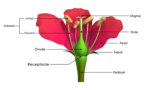
Free Images: "bestof:Anticlea elegans half longitudinal cut flower ovary ovules stamens style glands.svg es Anticlea elegans anteriormente Zigadenus elegans corte longitudinal de la"
Cyornis_turcosus_1838.jpg
Pulmonata.jpg
Haeckel_Calcispongiae.jpg
Layla_and_Majnun2.jpg
zinnia-elegans-zinnia-pink-flower-395612.jpg
zinnia-elegans-purple-flower-flower-1122897.jpg
zinnia-elegans-zinnia-pink-flower-404780.jpg
zinnia-elegans-zinnia-pink-flower-404773.jpg
zinnia-elegans-zinnia-pink-flower-403841.jpg
zinnia-elegans-zinnia-red-flowers-395613.jpg
zinnia-elegans-zinnia-pink-flower-398980.jpg
zinnia-zinnia-elegans-yellow-flowers-171292.jpg
poppy-gypsophila-elegans-red-color-1128683.jpg
anemone-vitifolia-anemone-elegans-876147.jpg
zinnia-daliowa-zinnia-elegans-zinnia-1273608.jpg
zinnia-daliowa-zinnia-elegans-zinnia-1273612.jpg
zinnia-daliowa-zinnia-elegans-zinnia-1273609.jpg
zinnia-daliowa-zinnia-elegans-zinnia-1273610.jpg
zinnia-daliowa-zinnia-elegans-zinnia-1273611.jpg
zinnia-daliowa-zinnia-elegans-zinnia-1273613.jpg
zinnia-daliowa-zinnia-elegans-zinnia-1273614.jpg
zinnia-daliowa-zinnia-elegans-zinnia-1273615.jpg
Qur'an_manuscript_Surat_al-Nisa'._(1).tif
marigold-zinnia-zinnia-elegans-123303.jpg
zinnia-zinnia-flower-zinnia-elegans-1480208.jpg
Melanopareia_elegans_elegans_1897.jpg
Bee on Zinnia elegans near Kings Park Eternal Flame-1-.jpg
zinnia-orange-zinnia-zinnia-elegans-1477228.jpg
zinnia-yellow-zinnia-zinnia-elegans-1477217.jpg
zinnia-pink-zinnia-zinnia-bloom-1477226.jpg
zinnia-flowers-colorful-color-59913.jpg
zinnia-flowers-colorful-color-59911.jpg
flowers-colorful-zinnia-color-59912.jpg
zinnia-flowers-colorful-color-59910.jpg
fig-fruit-ripe-227983.jpg
zinnia-flowers-red-color-colorful-64684.jpg
zinnia-flowers-red-color-colorful-64687.jpg
zinnia-blossom-bloom-colorful-61348.jpg
zinnia-blossom-bloom-colorful-61330.jpg
zinnia-blossom-bloom-colorful-61344.jpg
zinnia-blossom-bloom-colorful-61329.jpg
zinnia-blossom-bloom-colorful-61328.jpg
zinnia-blossom-bloom-colorful-61327.jpg
blossom-bloom-colorful-zinnia-61326.jpg
tibouchina-granulosa-glory-bush-1504763.jpg
Anemone,_Japanese.jpg
Araignées,_insectes_et_fleurs_de_la_forêt_de_Moulière_(Les_Agobis).jpg
Gartenpflanzen 2012-09-01-0296.jpg
Rose_de_Porcelaine.jpg
Casque Skanderbeg Vienne.jpg
Doña_Isabel_la_Católica_dictando_su_testamento_(Rosales).jpg
Ansozour_0001.jpg
Les_magnifiques_toxiques.jpg
Fac simile sorcier gravé.jpg
MRAH 271211 2 Seigneur et dame.jpg
MANNapoli 9058 couple painting.jpg
Garden_delights.jpg
Bleu_du_ciel,_sans_nuage.jpg
Griffin mosaic Rhodes museum.jpg
Elena Colosseo Rome Italy.jpg
Huerta_Orgánica_en_la_Embajada.jpg
Pistachier_térébinthe.jpg
Capitol Building Full View.jpg
Ecballium_elaterium.jpg
Fleurs_du_solstice.jpg
Eagle nebula pillars complete.jpg
Schloss Lembeck, 3.jpg
María_Cristina_de_Borbón-Dos_Sicilias,_reina_de_España.jpg
Mon_environnement.jpg
Pico Humboldt Venezuela cut.jpg
Kunyu_Wanguo_Quantu_(坤輿萬國全圖).jpg
Centranthus.jpg
Mon_environnement.jpg
Global tropical cyclone tracks-edit2.jpg
Pieter_Brueghel_the_Elder_-_The_Dutch_Proverbs_-_Google_Art_Project.jpg
Anticlea elegans half longitudinal cut flower ovary ovules stamens style glands.svg
Prunus longitudinal half cut flower ovary style stamens hypanthium.svg
Fragaria flower longitudinal section.svg
Flower morphology - longitudinal section chirimoya Annona.svg
Plant morphology - inflorescence asteraceae compositae longitudinal section.svg
Germ band blastoderm vitellophagous yolk in an insect egg.svg
Flower morphology - Flower longitudinal section bilabial Bignoniaceae Tecoma stans.svg
Respiratory horns on the egg of Nepa Hemiptera longitudinal section after Hinton 1961.svg
Flower morphology - longitudinal section showing achene formation and hypanthium in Rosa-01.svg
Fruit morphology - samara - flower longitudinal section wings on developing fruit Acer platanoides.svg
Sección_longitudinal_de_la_Iglesia_de_Torrero.jpg
Flower morphology - flower cut-away Ericaceae Arbutus.svg
Cerdido.svg
Ovule-Gymno-Angio-es.svg
Flower morphology perianth type carinate - parts keel.svg
Gynoecium morphology style geniculate.svg
Flower morphology - Flower lateral view some pieces removed - gynobasic style position epipetalous stamen Boraginaceae.svg
Gray769-es.svg
Plant morphology - inflorescence asteraceae compositae.svg
La_Infanta_María_Antonia_Fernanda,_hija_de_Felipe_V,_Jacopo_Amigoni.jpg
Chunnel traffic-fr.svg
Ranc_-_María_Ana_Victoria_de_Borbón.jpg
Sigurd.svg
Distribution of Russula elegans.svg
Juan_de_la_Corte_-_El_juicio_de_Salomón.JPG
Terms of Use   Search of the Day