Click Here for More Images from iStock- 15% off with coupon 15FREEIMAGES 
Mitochondria is a double membrane-bound organelle found in all eukaryotic organisms. are related to the cellular respiration process. 3D illustration
Particles, medical concept
Cell anatomy on scientific background. 3d illustration
3d Mitochondrion isolated on black
Bronchitis and alveolitis in lung of horse infected with equine influenza virus, 200x magnification.
Photomicrograph of filamentous blue-green algae, Oscillatoria species. Each band along the stand is one cell. This filament is very short probably having recently parted from a longer one. Filaments are typically many times longer. This algae is mobile, it moves and twists through the water. San Francisco Bay, California, USA. Live specimen. Wet mount, 40X objective, transmitted brightfield illumination. Note - motion blur of live specimen, very shallow depth of field, chromatic aberration and uneven focus are inherent in light microscopy.
Aerial view of loyster farm by Ardara, County Donegal - Ireland.
Abstract 3D illustration.
Licmophora sp. algae, marine and freshwater diatom under microscopic view. Genus of benthic, photosynthetic and epiphyte diatom
plant Flower bud of Lilium brownii var.viridulum T.S.\nunder light mircoscope with white background
Mitochondria, a membrane-enclosed cellular organelles producing energy, 3D illustration
Viral Infection Monkeypox Virus - 3d rendered image. Abstract biomedical illustration. \nAntibody, Antigen, Vaccine technology concept.
Cross-section view of Mitochondria, 3d rendering. Computer digital drawing.
Diffuse large B-cell lymphoma, or DLBCL, is a cancer that starts in white blood cells called lymphocytes.
Components of Eukaryotic cell, nucleus and organelles and plasma membrane - 3d illustration
Natural agate surfaces, suitable for backgrounds and wallpapers.
3d rendering of biological animal cell with organelles cross section isolated on white. Animal cell with placed text annotations to all organelles
Tetis of Fish T.S. under light microscope with white background
The intermembrane transporter of the Gram-negative bacterium Acinetobacter baylyi transports a lipopolysaccharide (orange) through the inner membrane consuming ATP (red)
Photomicrograph of flatworm,
Epidermis. Electron microscope micrograph showing a keratinocyte of spinous layer. The epithelial cell has a polygonal shape, central nucleus with nucleolus, cytoplasm full of keratin filament bundles, and numerous dark desmosomes crossing the intercellular spaces.
http://vamorelia.com/ibanners/7.jpg
Backgrounds of Characteristics Tissue Tongue, Small intestine of Cat and Tissue of Esophagus rabbit under microscope.
Microscopic image of dog stomach cardiac region
amazing inhabitants of the microworld under a microscope
plant rape flower cross section under light mircoscope with white background
Internal structure of an animal cell, 3d rendering. Section view. Computer digital drawing.
Myxoid liposarcoma.  Myxoid/round cell liposarcoma, or MRCLS, is one of several types of liposarcoma. Liposarcoma is a rare cancer that grows in the cells that store fat in the body. MRCLS usually grows in the arms and legs. These tumors grow slowly, and they can spread to other parts of the body
The most prominent roles of mitochondria are to produce the energy currency of the cell, ATP (i.e., phosphorylation of ADP), through respiration, and to regulate cellular metabolism.
Golden Turkish Lira Symbol
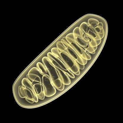
Free Images: "bestof:Animal mitochondrion diagram fr.svg mitochondrion mitochondrie File Animal mitochondrion diagram en svg 2006-05-02 translated by Ethan Gray original by"
Animal mitochondrion diagram fr.svg
Animal mitochondrion diagram hu.svg
Animal mitochondrion diagram hi.svg
Animal mitochondrion diagram it.svg
Animal mitochondrion diagram id.svg
Animal mitochondrion diagram ml.svg
Animal mitochondrion diagram zh hk.svg
Animal mitochondrion diagram zh tw.svg
Animal mitochondrion diagram zh ml.svg
Animal mitochondrion diagram fi.svg
Animal mitochondrion diagram pt.svg
Animal mitochondrion diagram pl.svg
Animal mitochondrion diagram de.svg
Animal mitochondrion diagram en.svg
Animal mitochondrion diagram uk.svg
Animal mitochondrion diagram en (edit).svg
Animal mitochondrion diagram ru.svg
Animal mitochondrion diagram cs.svg
Animal mitochondrion diagram es.svg
Animal mitochondrion diagram unlabelled.svg
Animal mitochondrion diagram lt.svg
Animal mitochondrion diagram ca.svg
Animal mitochondrion diagram bn.svg
Animal mitochondrion diagram gl.svg
Endomembrane system diagram en (edit).svg
Animal cell cycle-zh-hant.svg
Animal cell structure sv.svg
Animal cell structure id.svg
Acrosome reaction diagram es.svg
Mitochondrion_(borderless_version)-pt.svg
Animal Cell.svg
Animal cell structure no text.svg
Cell membrane detailed diagram id.svg
Animal cell structure fr.svg
Brain_diagram_ja.svg
Diagram of influenza infection symptoms EN.svg
Plant cell structure svg labels.svg
Mitochondrion mini.svg
PD Diagram of swine flu symptoms EN.svg
Mitochondrion (numbers version).svg
Animal cell cycle-en.svg
Animal cell structure gl.svg
Animal cell structure pl.svg
Animal cell cycle (editors version).svg
Animal cell structure en.svg
Gray code reflect.svg
VennDiagram.svg
Mitocondri (standalone version)-ca.svg
Mitocondri (borderless version)-ca.svg
Addresse Ipv4.svg
Animal cell structure de.svg
Hockey field metric fr.svg
Animal cell structure pt.svg
Average prokaryote cell- fr.svg
Endomembrane system diagram id.svg
Multipath propagation diagram de.svg
Digestive system diagram no labels arrows.svg
Complete neuron cell diagram he.svg
Escudo-de-la-Fe Simbolo-Atanasiano.svg
Plant cell wall diagram mk.svg
Tir a la corde equilibre.svg
Gray728.svg
Brain_diagram_pl.svg
Schematic diagram of the human eye ar.svg
Drupe fruit diagram-uk.svg
Leishmaniasis life cycle diagram ar.svg
Culex pipiens diagram fr.svg
Wikipedia article-creation-2 pl.svg
Endomembrane system diagram nl.svg
Endomembrane system diagram ln.svg
ะ›ะธะฝะตะฐั€ะตะฝ ะผะพั‚ะพั€.svg
Fluidized Bed Reactor Graphic-ru.svg
Cloud computing-id.svg
Complete diagram of a human spermatozoa pt.svg
Diagram human cell nucleus oc.svg
Wikipedia article-creation-2 sq.svg
Temporo-parietal_junction_ja.svg
Deltavs-fr.svg
16CygBbOrbit-ca.svg
Leishmaniasis life cycle diagram ru.svg
Genetic code mk.svg
Average prokaryote cell- lt.svg
Human skeleton back sl.svg
Axial skeleton diagram pt.svg
Solid oxide fuel cell protonic mk.svg
Diagram of swine influenza symptoms EN.svg
Complete diagram of a human spermatozoa pl.svg
Circulatory System en edited.svg
Human skeleton front sl.svg
Culex pipiens diagram zh.svg
Homology vertebrates-fr.svg
Turgor pressure on plant cells diagram (zh-tw).svg
Punnett square mendel flowers mk.svg
Esquema de la batalla de Watling Street.svg
Circulatory System ru.svg
Prokaryote cell-ro.svg
Elementary particle interactions (is).svg
Average prokaryote cell- nl.svg
Average prokaryote cell- id.svg
Elementary particle interactions (polish version) pl.svg
Terms of Use   Search of the Day