Click Here for More Images from iStock- 15% off with coupon 15FREEIMAGES 
Internal structure of an animal cell, 3d rendering. Section view. Computer digital drawing.
Hepatocellular adenoma (HCA; also termed hepatic adenoma) is an uncommon solid, benign liver lesion that develops in an otherwise normal-appearing liver. Typically, HCAs are solitary and are found in young women in association with use of estrogen-containing medications.
brown, beige and white fat cells,  adipocyte and lipocyte, cholesterol in a cells, adipose tissue, lipid droplet, \nfat in body, Obesity, Types of lipocytes dermis and hypodermis, nucleus, 3d render
3d Mitochondrion isolated on black
Bronchitis and alveolitis in lung of horse infected with equine influenza virus, 200x magnification.
Photomicrograph of filamentous blue-green algae, Oscillatoria species. Each band along the stand is one cell. This filament is very short probably having recently parted from a longer one. Filaments are typically many times longer. This algae is mobile, it moves and twists through the water. San Francisco Bay, California, USA. Live specimen. Wet mount, 40X objective, transmitted brightfield illumination. Note - motion blur of live specimen, very shallow depth of field, chromatic aberration and uneven focus are inherent in light microscopy.
Illustration of animal cell anatomy
Abstract 3D illustration.
centropyxis aculeata (amoeboid organism) under the microscope - optical microscope x400 magnification
plant rape flower cross section under light mircoscope with white background
Mitochondria, a membrane-enclosed cellular organelles producing energy, 3D illustration
Particles, medical concept
3d rendering of biological animal cell with organelles cross section isolated on white. Animal cell with placed text annotations to all organelles
Natural agate surfaces, suitable for backgrounds and wallpapers.
Internal structure of an animal cell, 3d rendering. Section view. Computer digital drawing.
Fatty liver disease (steatosis) is a common condition caused by having too much fat build up in your liver. A healthy liver contains a small amount of fat. It becomes a problem when fat reaches 5% to 10% of your liver's weight.
Leiden leaf c.s. under microscope
plant Stem-Collenchyma C.S. under light mircoscope with white background
amazing inhabitants of the microworld under a microscope
Photomicrograph of two rotifers. Rapidly rotating cilia at top of heads, internal organs visible. Live specimen. Wet mount, 10X objective, transmitted brightfield illumination.
Aerial view of loyster farm by Ardara, County Donegal - Ireland.
Squash preparation of  onion root tip cells stained to reveal chromosomes,  Bright field illumination, light microscopy.  This image was taken with the X40 objective lens.  Depth of focus is extremely low at this magnification.  Smaller apertures give greater depth of focus, but image quality deteriorates and resolution is greatly reduced.  The aperture in this image was optimised for contrast and resolution. Chromatic abberation is inevitable in light microscopy, but has been kept to a minimum in this image.
Freshwater Closterium algae (unicellular charophyte green algae) - optical microscope x200 magnification
Microscopic image of dog stomach cardiac region
Licmophora sp. algae, marine and freshwater diatom under microscopic view. Genus of benthic, photosynthetic and epiphyte diatom
Tetis of Fish T.S. under light microscope with white background
Cells consist of a protoplasm enclosed within a membrane, which contains many biomolecules such as proteins and nucleic acids.
Pheochromocytoma, Site: Adrenal gland
Micrograph of rat brain. Science cross section.
Curly marble stone with fashion veins.
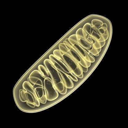
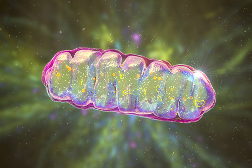
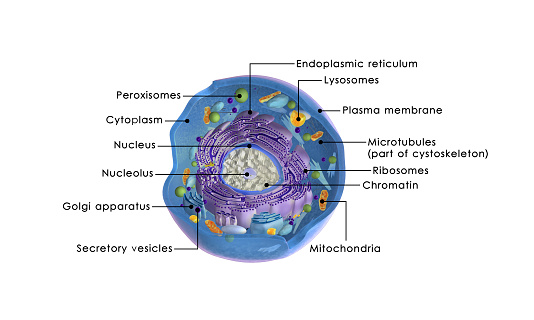
Free Images: "bestof:Animal mitochondrion diagram fi.svg fi mitokondrio File Animal mitochondrion diagram en svg 2007-05-06 translated by B Nuhanen based on image by Mariana Ruiz"
Animal mitochondrion diagram fi.svg
Animal mitochondrion diagram pl.svg
Animal mitochondrion diagram pt.svg
Animal mitochondrion diagram hu.svg
Animal mitochondrion diagram id.svg
Animal mitochondrion diagram zh hk.svg
Animal mitochondrion diagram zh tw.svg
Animal mitochondrion diagram ml.svg
Animal mitochondrion diagram zh ml.svg
Animal mitochondrion diagram ru.svg
Animal mitochondrion diagram it.svg
Animal mitochondrion diagram es.svg
Animal mitochondrion diagram uk.svg
Animal cell structure id.svg
Animal cell structure sv.svg
Animal mitochondrion diagram en.svg
Animal mitochondrion diagram en (edit).svg
Animal mitochondrion diagram cs.svg
Animal mitochondrion diagram fr.svg
Animal mitochondrion diagram hi.svg
Animal cell structure de.svg
Animal cell structure no text.svg
Animal cell structure en.svg
Gap cell junction it.svg
Cell junction simplified it.svg
Animal mitochondrion diagram bn.svg
Cell junction simplified pt.svg
Animal mitochondrion diagram de.svg
Animal mitochondrion diagram lt.svg
Animal mitochondrion diagram unlabelled.svg
Adherens Junctions structural proteins.svg
Animal cell structure pl.svg
Endomembrane system diagram en.svg
Bacterial morphology diagram cs.svg
Animal cell structure pt.svg
Diagram human cell nucleus sl.svg
Endomembrane system diagram cs.svg
Diagram human cell nucleus cs.svg
Endomembrane system diagram id.svg
Prokaryote cell diagram cs.svg
Cell membrane detailed diagram id.svg
Diagram human cell nucleus id.svg
Leishmaniasis life cycle diagram ar.svg
Endomembrane system diagram ka.svg
Leishmaniasis life cycle diagram ru.svg
Human skeleton back sl.svg
Circulatory System en edited.svg
Culex pipiens diagram uk.svg
Human skeleton front sl.svg
Culex pipiens diagram fr.svg
Complete diagram of a human spermatozoa pt.svg
Endomembrane system diagram nl.svg
Endomembrane system diagram ln.svg
PD Diagram of swine flu symptoms EN.svg
Circulatory System ru.svg
Animal mitochondrion diagram gl.svg
Culex pipiens diagram es.svg
Animal mitochondrion diagram ca.svg
Diagram of influenza infection symptoms EN.svg
Average prokaryote cell- id.svg
Diagram of swine influenza symptoms EN.svg
Complete diagram of a human spermatozoa pl.svg
Adherens Junctions structural proteins-fr.svg
Plant cell structure svg labels.svg
Prokaryote cell-ro.svg
Average prokaryote cell- nl.svg
Digestive system diagram fi.svg
Acrosome reaction diagram es.svg
Morphologie bei Bakterien.svg
Mature flower diagram-lmo.svg
Competitive inhibitor Fi.svg
Wikipedia article creation fi.svg
Competitive inhibitor.svg
Digestive system diagram sr.svg
Endomembrane system diagram fr.svg
Flagellum base diagram keys.svg
Digestive system diagram mk.svg
Prokaryote cell diagram pt.svg
Prokaryote cell diagram no.svg
Wine grape diagram en.svg
Endomembrane system diagram en (edit).svg
Simplified spermatozoon diagram cs.svg
Average prokaryote cell- fr.svg
Animal cell cycle-zh-hant.svg
Diagram human cell nucleus tr.svg
Diagram human cell nucleus pt.svg
Cross section jellyfish en.svg
Gap cell junction-cs.svg
Prokaryote cell diagram he.svg
Mendel seven characters cs.svg
Lebenszyklus der Angiospermen.svg
Filtration diagram (zh-tw).svg
Connexon and connexin structure cs.svg
Cell junction simplified cs.svg
Diagram human cell nucleus hu.svg
Human skull side suturas pl.svg
Gap cell junction-de.svg
16CygBbOrbit-ca.svg
Cell junction simplified zh traditional.svg
Digestive system diagram no labels arrows.svg
Terms of Use   Search of the Day